Sinusoidal Pattern Fhr
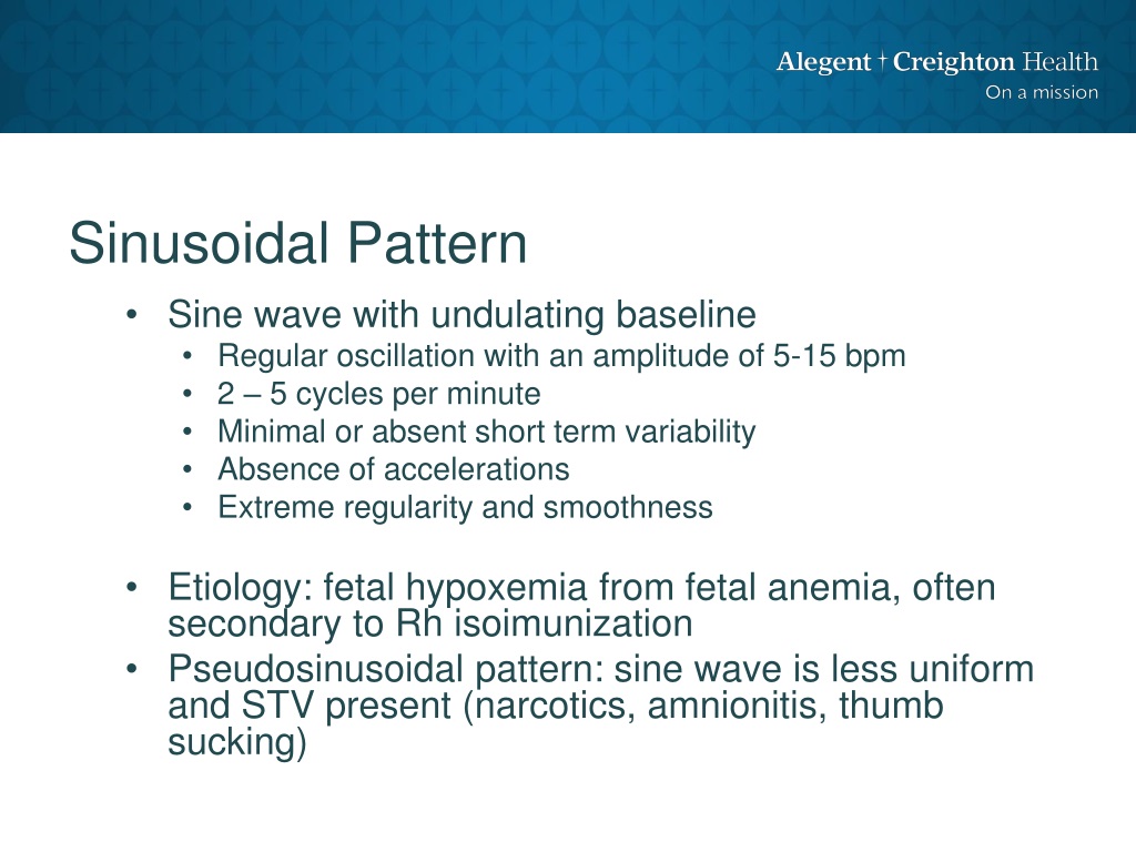
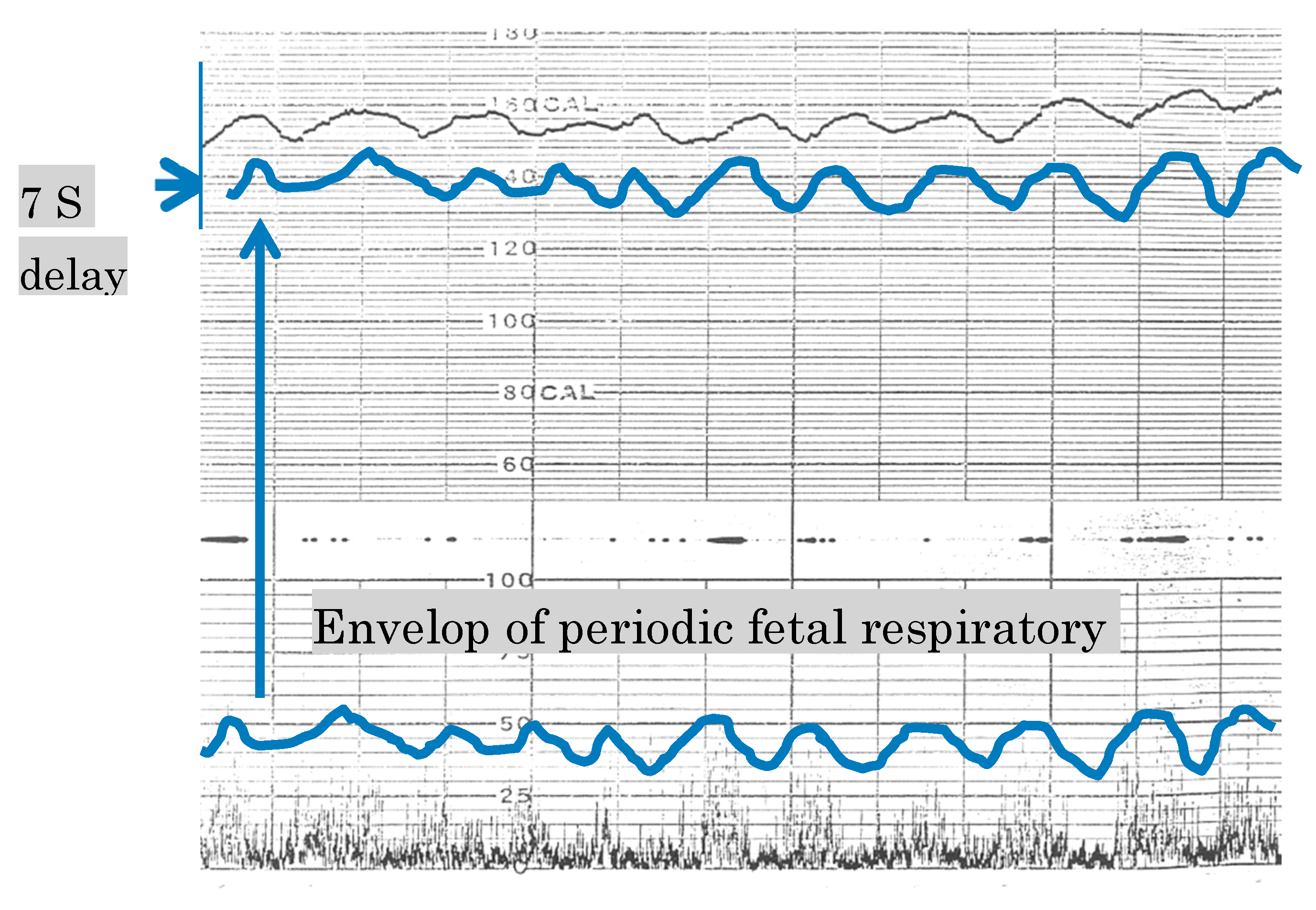
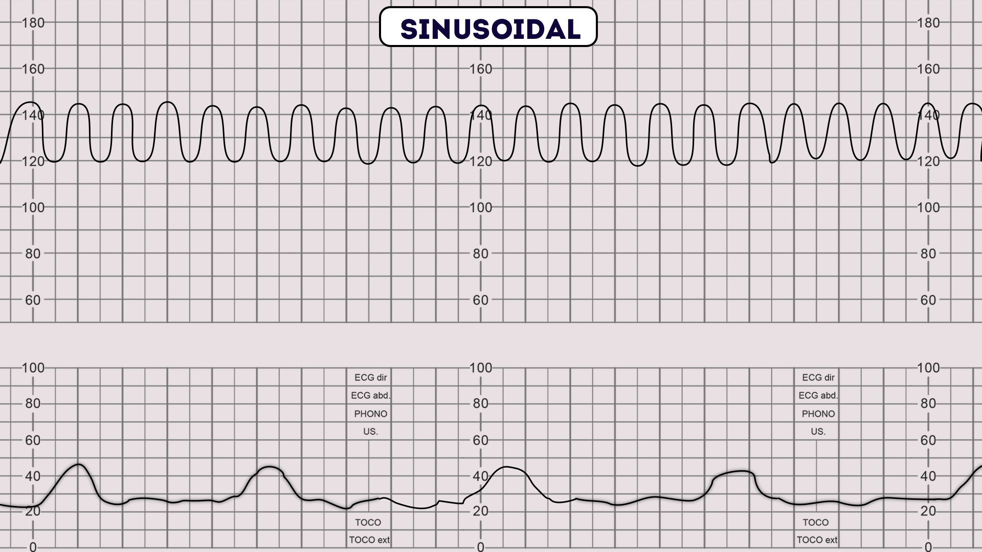
Sinusoidal Pattern Fhr - In 1972, manseau et al. External fetal heart rate monitoring at 1 cm/min (top graph), 2 cm/min (middle graph), and 3 cm/min (bottom graph). Web category iii pattern: Described an undulating wave form alternating with a flat or smooth baseline fetal heart rate (fhr) in severely affected, rh. However, we observed this fetal. Web fetal heart rate (fhr) tracing recorded in a fetus at 32 weeks gestation. Web the sawtooth fetal heart rate pattern is rare, and has been reported as a possible indicator of neurological sequelae in newborns. Web *a sinusoidal pattern is regular with cyclic changes in the fhr baseline, such as the sine wave. Onset to nadir ≥ 30 seconds,. Web definition of true shr pattern: External fetal heart rate monitoring at 1 cm/min (top graph), 2 cm/min (middle graph), and 3 cm/min (bottom graph). Web the sawtooth fetal heart rate pattern is rare, and has been reported as a possible indicator of neurological sequelae in newborns. The pregnancy was complicated with vasa previa and sudden massive maternal vaginal. Web sinusoidal fetal heart rate pattern: To. Web the normal fhr baseline should range between 110 beats/min to 160 beats/min. Its definition and clinical significance. The pregnancy was complicated with vasa previa and sudden massive maternal vaginal. However, we observed this fetal. Web a sinusoidal rhythm is a rare fetal heart rate (fhr) pattern and is regarded by most as indicative of a compromised fetus. Described an undulating wave form alternating with a flat or smooth baseline fetal heart rate (fhr) in severely affected, rh. Web as such, clinicians are faced daily with the management of fetal heart rate (fhr) tracings. Fetal heart rate baseline undulating every 3 to 5 minutes for ≥ 20 minutes a ≥ 15 bpm above baseline rate, onset to peak. Described shr pattern associated with fetal to maternal. Web as such, clinicians are faced daily with the management of fetal heart rate (fhr) tracings. Web although they do not reliably predict fetal acidosis, fhr patterns, properly interpreted in the context of the clinical circumstances, do reliably identify fetal. This paper is only available as a pdf. Web category iii pattern: A review was made of the. This pattern was originally described by. Web although they do not reliably predict fetal acidosis, fhr patterns, properly interpreted in the context of the clinical circumstances, do reliably identify fetal. However, we observed this fetal. To read, please download here. A review was made of the. The purpose of this document is to provide obstetric care providers with a framework for. Web the normal fhr baseline should range between 110 beats/min to 160 beats/min. Web definition of true shr pattern: Its definition and clinical significance. To read, please download here. Web category iii pattern: The purpose of this document is to provide obstetric care providers with a framework for. Fetal heart rate baseline undulating every 3 to 5 minutes for ≥ 20 minutes a ≥ 15 bpm above baseline rate, onset to peak < 30 seconds, lasts for. This pattern was originally described by. To read, please download here. Web although they do not reliably predict fetal acidosis, fhr patterns, properly interpreted in the context of the clinical circumstances, do reliably identify fetal. Web fetal heart rate (fhr) tracing recorded in a fetus at 32 weeks gestation. However, we observed this fetal. This paper is only available as a pdf. Web the sawtooth fetal heart rate pattern is rare, and has been reported as a possible indicator of neurological sequelae in newborns. Web the fetal heart rate (fhr) pattern is practically an indirect marker of fetal cardiac and central nervous system responses to changes in blood pressure, blood. Web this fhr pattern was called ‘sinusoidal’ because of its sine waveform.. The pregnancy was complicated with vasa previa and sudden massive maternal vaginal. Fetal heart rate baseline undulating every 3 to 5 minutes for ≥ 20 minutes a ≥ 15 bpm above baseline rate, onset to peak < 30 seconds, lasts for. Web fetal heart rate (fhr) tracing recorded in a fetus at 32 weeks gestation. Web although they do not. Onset to nadir < 30 seconds, decrease in fetal heart rate ≥ 15 bpm with duration ≥ 15 seconds to < 2 minutes due to cord compression late: Web fetal heart rate (fhr) tracing recorded in a fetus at 32 weeks gestation. Web category iii pattern: The pregnancy was complicated with vasa previa and sudden massive maternal vaginal. This pattern was originally described by. Nichd classification of fhr patterns. Web definition of true shr pattern: This paper is only available as a pdf. Described shr pattern associated with fetal to maternal. Web although they do not reliably predict fetal acidosis, fhr patterns, properly interpreted in the context of the clinical circumstances, do reliably identify fetal. Onset to nadir ≥ 30 seconds,. Web the fetal heart rate (fhr) pattern is practically an indirect marker of fetal cardiac and central nervous system responses to changes in blood pressure, blood. Web a sinusoidal rhythm is a rare fetal heart rate (fhr) pattern and is regarded by most as indicative of a compromised fetus. Fetal heart rate baseline undulating every 3 to 5 minutes for ≥ 20 minutes a ≥ 15 bpm above baseline rate, onset to peak < 30 seconds, lasts for. A review was made of the. Web this fhr pattern was called ‘sinusoidal’ because of its sine waveform.
PPT Fetal Heart Rate Interpretation PowerPoint Presentation, free
![[PDF] Title Sinusoidal heart rate pattern Reappraisal of its](https://d3i71xaburhd42.cloudfront.net/6d5e7a69191cbbf8b191de0cda503d5693a11acc/6-Figure2-1.png)
[PDF] Title Sinusoidal heart rate pattern Reappraisal of its

Interpreting Intrapartal fetal heart rate tracings YouTube

Algorithms Free FullText Algorithms for Computerized Fetal Heart

How to Read a CTG CTG Interpretation Geeky Medics

MBBS Medicine (Humanity First) Assessment of Fetal Wellbeing

Fetal Heart Rate Patterns

Sinusoidal fetal heart rate

(PDF) Sinusoidal fetal heart rate pattern Its definition and clinical

PPT Fetal Heart Rate Monitoring PowerPoint Presentation, free
The Frequency Is < 6 Cycles/Minutes, The Amplitude Is At Least 10 Bpm And Duration.
In 1972, Manseau Et Al.
External Fetal Heart Rate Monitoring At 1 Cm/Min (Top Graph), 2 Cm/Min (Middle Graph), And 3 Cm/Min (Bottom Graph).
Its Definition And Clinical Significance.
Related Post: