Sinusoidal Pattern Ecg
Sinusoidal Pattern Ecg - (see normal sinus rhythm and sinus arrhythmia.) because of this activation. A standard 12 lead ecg is shown in [figure 1]. Serum potassium level may not correlate closely with ecg changes. This is possible because there is a correlation between the severity of electrolyte imbalance and the visible ecg changes. Sinus rhythm is present when the dominant pacemaker controlling impulse generation is the sinus node ( waveform 1 and waveform 2 ). In this setting, activation of the atria is from right to left, superior to inferior, and anterior to posterior. Web the ecg may be used to estimate the severity of electrolyte imbalances and to judge whether there is a risk of serious arrhythmias. The best way to interpret an ecg is to read it systematically: The pr interval remains constant. Sinoatrial node (sa node) atrioventricular node (av node) his bundle left and right bundle branches purkinje fibers myocytes. The best way to interpret an ecg is to read it systematically: P waves upright in leads i and ii, inverted in avr. Test results can help diagnose heart attacks and irregular heartbeats, called arrhythmias. Sinus rhythm is present when the dominant pacemaker controlling impulse generation is the sinus node ( waveform 1 and waveform 2 ). An action potential. An action potential leads to myocyte depolarization, which spreads quickly among adjoining cells. Test results can help diagnose heart attacks and irregular heartbeats, called arrhythmias. Web ecg features of normal sinus rhythm. A standard 12 lead ecg is shown in [figure 1]. Web sinus rhythm is the normal rhythm of the heart and no treatment is relevant. In this setting, activation of the atria is from right to left, superior to inferior, and anterior to posterior. Serum potassium level may not correlate closely with ecg changes. The best way to interpret an ecg is to read it systematically: Arrhythmias from the sinoatrial node are also discussed. Learn the definition, physiology, criteria for sinus rhythm, with emphasis on. Sinus rhythm is present when the dominant pacemaker controlling impulse generation is the sinus node ( waveform 1 and waveform 2 ). A standard 12 lead ecg is shown in [figure 1]. Electrical signal interpretation gives a good approximation of heart pathology. Patients with a relatively normal ecg can suffer sudden hyperkalaemia cardiac arrest. Web sinus rhythm is the normal. The best way to interpret an ecg is to read it systematically: Web ecg features of normal sinus rhythm. This is possible because there is a correlation between the severity of electrolyte imbalance and the visible ecg changes. (see normal sinus rhythm and sinus arrhythmia.) because of this activation. Arrhythmias from the sinoatrial node are also discussed. It records the electrical signals in the heart. In this setting, activation of the atria is from right to left, superior to inferior, and anterior to posterior. Arrhythmias from the sinoatrial node are also discussed. (see normal sinus rhythm and sinus arrhythmia.) because of this activation. Pea with bizarre, wide complex rhythm; Web the ecg may be used to estimate the severity of electrolyte imbalances and to judge whether there is a risk of serious arrhythmias. P waves upright in leads i and ii, inverted in avr. It records the electrical signals in the heart. Test results can help diagnose heart attacks and irregular heartbeats, called arrhythmias. The best way to interpret. Sinus rhythm is present when the dominant pacemaker controlling impulse generation is the sinus node ( waveform 1 and waveform 2 ). This is possible because there is a correlation between the severity of electrolyte imbalance and the visible ecg changes. Arrhythmias from the sinoatrial node are also discussed. Web sinus rhythm is the normal rhythm of the heart and. (see normal sinus rhythm and sinus arrhythmia.) because of this activation. P waves upright in leads i and ii, inverted in avr. Patients with a relatively normal ecg can suffer sudden hyperkalaemia cardiac arrest. The best way to interpret an ecg is to read it systematically: Sinoatrial node (sa node) atrioventricular node (av node) his bundle left and right bundle. Web the goal of the electrocardiogram interpretation is to determine whether the ecg waves and intervals are normal or pathological. In this setting, activation of the atria is from right to left, superior to inferior, and anterior to posterior. Pea with bizarre, wide complex rhythm; Sinoatrial node (sa node) atrioventricular node (av node) his bundle left and right bundle branches. P waves upright in leads i and ii, inverted in avr. In this setting, activation of the atria is from right to left, superior to inferior, and anterior to posterior. Pea with bizarre, wide complex rhythm; Web ecg features of normal sinus rhythm. Serum potassium level may not correlate closely with ecg changes. Electrical signal interpretation gives a good approximation of heart pathology. The best way to interpret an ecg is to read it systematically: Sinus rhythm is present when the dominant pacemaker controlling impulse generation is the sinus node ( waveform 1 and waveform 2 ). Web an electrocardiogram (ecg or ekg) is a quick test to check the heartbeat. Web the ecg may be used to estimate the severity of electrolyte imbalances and to judge whether there is a risk of serious arrhythmias. An action potential leads to myocyte depolarization, which spreads quickly among adjoining cells. Learn the definition, physiology, criteria for sinus rhythm, with emphasis on ecg interpretation. Patients with a relatively normal ecg can suffer sudden hyperkalaemia cardiac arrest. The pr interval remains constant. Test results can help diagnose heart attacks and irregular heartbeats, called arrhythmias. Sinoatrial node (sa node) atrioventricular node (av node) his bundle left and right bundle branches purkinje fibers myocytes.
Pin on internal medicine

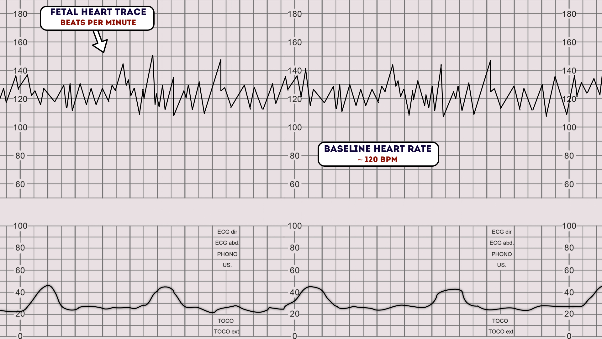
Sinusoidal fetal heart rate

MBBS Medicine (Humanity First) Assessment of Fetal Wellbeing

ECG changes due to electrolyte imbalance (disorder) ECG & ECHO

Patrón de onda sinusoidal TJMBB

Understanding the EKG Signal Atrial Fibrillation Resources for Patients

Sine Wave Pattern Ecg Images and Photos finder

Ecg sinusoidal pulse lines frequency heartbeat Vector Image

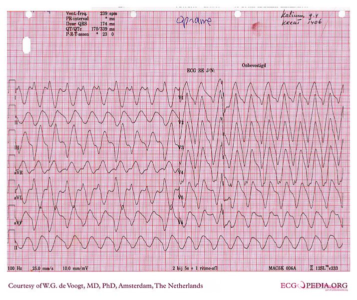
12 lead EKG showing sinewave done in the emergency room. Download

Dr. Smith's ECG Blog Weakness and Dyspnea with a Sine Wave. It's not
This Is Possible Because There Is A Correlation Between The Severity Of Electrolyte Imbalance And The Visible Ecg Changes.
(See Normal Sinus Rhythm And Sinus Arrhythmia.) Because Of This Activation.
A Standard 12 Lead Ecg Is Shown In [Figure 1].
Web Sinus Rhythm Is The Normal Rhythm Of The Heart And No Treatment Is Relevant.
Related Post: