Sine Wave Pattern Ecg
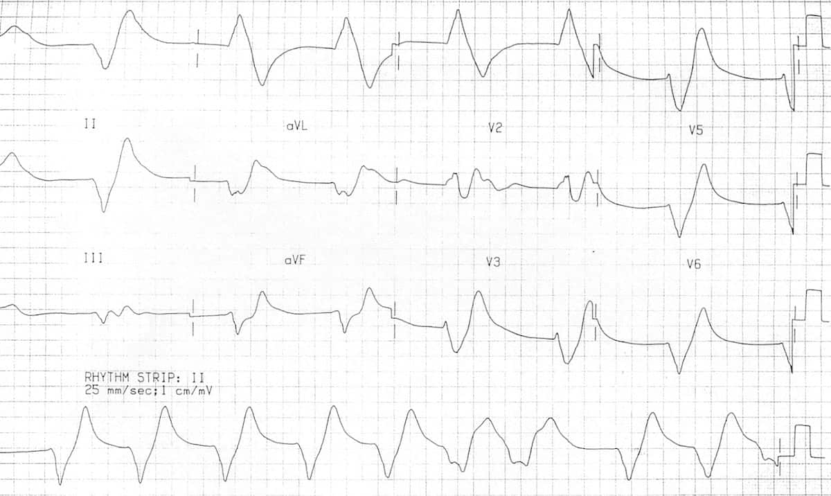
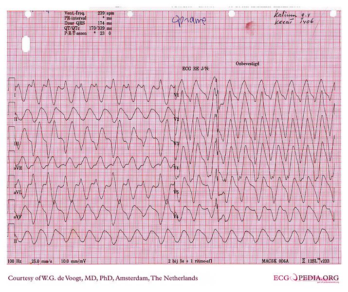
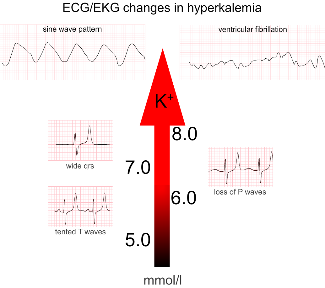
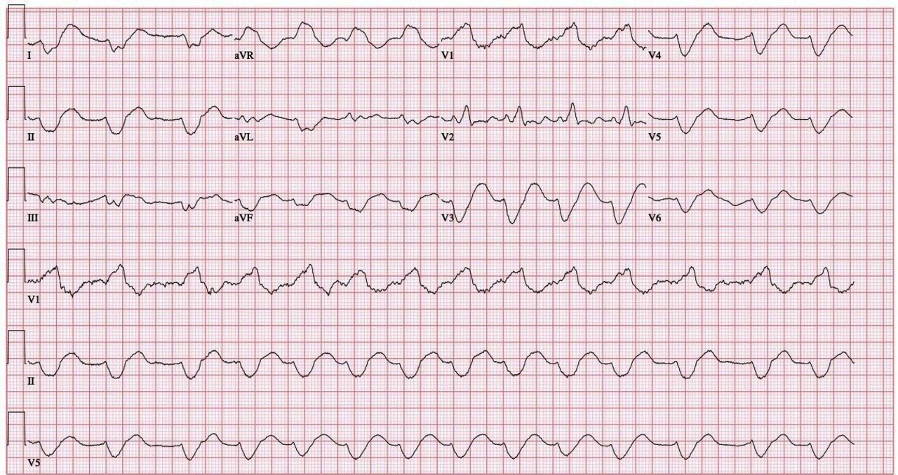
Sine Wave Pattern Ecg - Ecg changes begin with peaked t waves and widening p wave, and progress to sine wave appearance with severe hyperkalaemia. This article is an answer to the ecg case 174. Sinoatrial node (sa node) atrioventricular node (av node) his bundle left and right bundle branches purkinje. Web it involves looking for five points: The only condition that will prolonged the qt ≥ 0.24 sec is hyperkalemia, as a. Had we seen the earlier. Thyroid, high left ventricular voltage, hlvv, ecg diagnosis: Aacn adv crit care (2015) 26 (2): The sine wave pattern depicts worsening cardiac conduction delay caused by the elevated. Web severe hyperkalemia with sine wave ecg pattern. The t waves (+) are symmetric, although not tall or peaked. Ecg changes begin with peaked t waves and widening p wave, and progress to sine wave appearance with severe hyperkalaemia. Had we seen the earlier. 1 potassium was 8.4 mmol/l and. Thyroid, high left ventricular voltage, hlvv, ecg diagnosis: The t waves (+) are symmetric, although not tall or peaked. The web page explains the pathophysiology, causes, symptoms. Web ecg changes due to hypercalcemia. Web ecg showed pacemaker spikes followed by widened qrs complexes fused with tall t waves, similar to a sine wave pattern ( figure 2 a). The physical examination was unremarkable,. Web it involves looking for five points: The only condition that will prolonged the qt ≥ 0.24 sec is hyperkalemia, as a. Hyperkalaemia is defined as plasma potassium ≥ 5.5 mmol/l. The sine wave pattern depicts worsening cardiac conduction delay caused by the elevated. The patient was treated with 2 g of intravenous calcium gluconate, nebulised albuterol treatments, 10 units. Ecg changes begin with peaked t waves and widening p wave, and progress to sine wave appearance with severe hyperkalaemia. Changes need to occur in at least 2 of the right precordial leads (v1. Severe hyperkalemia may be devastating to. Web ecg changes due to hypercalcemia. Thyroid, high left ventricular voltage, hlvv, ecg diagnosis: Web it involves looking for five points: The presence or absence of regular p waves, the duration of qrs complexes (narrow or wide), the correlation between p waves and qrs. Web hyperkalemia with sine wave pattern. The t waves (+) are symmetric, although not tall or peaked. Web sine wave electrocardiogram (ecg) pattern in hyperkalemia is still a medical curiosity. The only condition that will prolonged the qt ≥ 0.24 sec is hyperkalemia, as a. Hyperkalemia (serum potassium > 5.5 meq/l [> 5.5. Web it involves looking for five points: The physical examination was unremarkable,. Web hyperkalaemia is a serum potassium level of > 5.2 mmol/l. Sinoatrial node (sa node) atrioventricular node (av node) his bundle left and right bundle branches purkinje. The patient was treated with 2 g of intravenous calcium gluconate, nebulised albuterol treatments, 10 units of regular insulin. This pattern usually appears when the serum potassium levels are well over 8.0 meq/l. The web page explains the pathophysiology, causes, symptoms. Web it involves. Aacn adv crit care (2015) 26 (2): The web page explains the pathophysiology, causes, symptoms. Web hyperkalemia with sine wave pattern. Had we seen the earlier. This article is an answer to the ecg case 174. Severe hyperkalemia may be devastating to. Web ecg showed pacemaker spikes followed by widened qrs complexes fused with tall t waves, similar to a sine wave pattern ( figure 2 a). The t waves (+) are symmetric, although not tall or peaked. Sinoatrial node (sa node) atrioventricular node (av node) his bundle left and right bundle branches purkinje. Aacn adv. Web hyperkalaemia is a serum potassium level of > 5.2 mmol/l. Ecg changes begin with peaked t waves and widening p wave, and progress to sine wave appearance with severe hyperkalaemia. The sine wave pattern is one of the manifestations of severe hyperkalemia. Web sine wave electrocardiogram (ecg) pattern in hyperkalemia is still a medical curiosity and rare to observe. Web it involves looking for five points: Hyperkalemia (serum potassium > 5.5 meq/l [> 5.5. The physical examination was unremarkable,. Web hyperkalemia with sine wave pattern. Severe hyperkalemia may be devastating to. Web ecg showed pacemaker spikes followed by widened qrs complexes fused with tall t waves, similar to a sine wave pattern ( figure 2 a). Web severe hyperkalemia with sine wave ecg pattern. Aacn adv crit care (2015) 26 (2): Sinoatrial node (sa node) atrioventricular node (av node) his bundle left and right bundle branches purkinje. 1 potassium was 8.4 mmol/l and. The t waves (+) are symmetric, although not tall or peaked. This article is an answer to the ecg case 174. Hyperkalaemia is further classified by the european resuscitation guidelines as follows:. The sine wave pattern depicts worsening cardiac conduction delay caused by the elevated. Thyroid, high left ventricular voltage, hlvv, ecg diagnosis: The sine wave pattern is one of the manifestations of severe hyperkalemia.
Sine Wave Hyperkalemia Ecg Changes

ECG changes due to electrolyte imbalance (disorder) ECG & ECHO

12 lead EKG showing sinewave done in the emergency room. Download

Ecg sinusoidal pulse lines frequency heartbeat Vector Image

Hyperkalaemia ECG changes • LITFL • ECG Library

Sine wave pattern wikidoc

sine wave ecg

Hyperkalemia ecg findings garetbetter

Sinewave electrocardiogram rhythm in a patient on haemodialysis

Sine Wave Pattern Ecg Images and Photos finder
The Web Page Explains The Pathophysiology, Causes, Symptoms.
Web Ecg Changes Due To Hypercalcemia.
Thyroid, High Left Ventricular Voltage, Hlvv, Ecg Diagnosis:
Web Sine Wave Electrocardiogram (Ecg) Pattern In Hyperkalemia Is Still A Medical Curiosity And Rare To Observe In Routine Clinical Practice.
Related Post: