Rv Strain Pattern
Rv Strain Pattern - Pattern 2 was characterized by. Right heart strain can be caused by pulmonary hypertension, pulmonary embolism (or pe, which itself can cause pulmonary hypertension ), rv infarction (a heart attack affecting the rv), chronic lung disease (such as pulmonary fibrosis Web the three main parameters of rv strain are right ventricular global longitudinal strain (rvgls), right ventricular free wall longitudinal strain (rvfwls). Web echocardiographically, those with events had decreased free wall and global rv strain in addition to a larger left atrial volume index, more severe secondary mitral. Web in the last years, rv strain imaging emerged as a superior metric of rv systolic performance, overcoming some of the limitations of conventional. Web among various imaging modalities, echocardiography is currently the method of choice for clinical assessment of rv longitudinal strain (rvls). Find and treat the cause (e.g. Web in their study, a considerable proportion of patients with normal fractional area change and normal tricuspid annulus systolic excursion had abnormal rv longitudinal strain,. Web right ventricular strain pattern due to rvh: Web diagnosis of pe and right ventricular (rv) strain with transthoracic echocardiography (tte) however, has been well documented as a predictor for pending. Web highly trained athletes, especially those involved in endurance sport disciplines, can develop marked right ventricular (rv) remodeling that could raise the. Web right heart strain (or more precisely right ventricular strain) is a term given to denote the presence of right ventricular dysfunction usually in the absence of an. Web in their study, a considerable proportion of patients with. Clinical echocardiography miscellaneous conditions right ventricular strain: It is not known, however, if rv strain occurs in the. Web among various imaging modalities, echocardiography is currently the method of choice for clinical assessment of rv longitudinal strain (rvls). Web the three main parameters of rv strain are right ventricular global longitudinal strain (rvgls), right ventricular free wall longitudinal strain (rvfwls).. Web in the last years, rv strain imaging emerged as a superior metric of rv systolic performance, overcoming some of the limitations of conventional. Web in their study, a considerable proportion of patients with normal fractional area change and normal tricuspid annulus systolic excursion had abnormal rv longitudinal strain,. Web the three main parameters of rv strain are right ventricular. Right heart strain (also right ventricular strain or rv strain) is a medical finding of right ventricular dysfunction where the heart muscle of the right ventricle (rv) is deformed. Web right heart strain (or more precisely right ventricular strain) is a term given to denote the presence of right ventricular dysfunction usually in the absence of an. Web in their. Web rv strain was defined as in the presence of one or more of the following ecg findings: Web in their study, a considerable proportion of patients with normal fractional area change and normal tricuspid annulus systolic excursion had abnormal rv longitudinal strain,. Web the three main parameters of rv strain are right ventricular global longitudinal strain (rvgls), right ventricular. Web highly trained athletes, especially those involved in endurance sport disciplines, can develop marked right ventricular (rv) remodeling that could raise the. It is not known, however, if rv strain occurs in the. Web echocardiographically, those with events had decreased free wall and global rv strain in addition to a larger left atrial volume index, more severe secondary mitral. Pattern. Web rv strain was defined as in the presence of one or more of the following ecg findings: Learn how to diagnose and manage rvh with ecg review, criteria, and. Other features of rvh are present, including right axis deviation, and a dominant r wave in v1 Web diagnosis of pe and right ventricular (rv) strain with transthoracic echocardiography (tte). Web echocardiographically, those with events had decreased free wall and global rv strain in addition to a larger left atrial volume index, more severe secondary mitral. Right heart strain (also right ventricular strain or rv strain) is a medical finding of right ventricular dysfunction where the heart muscle of the right ventricle (rv) is deformed. Web highly trained athletes, especially. It is not known, however, if rv strain occurs in the. Web in their study, a considerable proportion of patients with normal fractional area change and normal tricuspid annulus systolic excursion had abnormal rv longitudinal strain,. Web right ventricular hypertrophy (rvh) is a condition that affects the heart's structure and function. Web among various imaging modalities, echocardiography is currently the. Right heart strain (also right ventricular strain or rv strain) is a medical finding of right ventricular dysfunction where the heart muscle of the right ventricle (rv) is deformed. Web rv strain was defined as in the presence of one or more of the following ecg findings: Web echocardiographically, those with events had decreased free wall and global rv strain. Clinical echocardiography miscellaneous conditions right ventricular strain: Web right heart strain (or more precisely right ventricular strain) is a term given to denote the presence of right ventricular dysfunction usually in the absence of an. Right heart strain (also right ventricular strain or rv strain) is a medical finding of right ventricular dysfunction where the heart muscle of the right ventricle (rv) is deformed. Other features of rvh are present, including right axis deviation, and a dominant r wave in v1 Web right ventricular strain pattern due to rvh: Web echocardiographically, those with events had decreased free wall and global rv strain in addition to a larger left atrial volume index, more severe secondary mitral. Web right ventricular hypertrophy (rvh) is a condition that affects the heart's structure and function. Pe, ards, mv failure, fluid overload, pulmonary htn) management focuses on: Web diagnosis of pe and right ventricular (rv) strain with transthoracic echocardiography (tte) however, has been well documented as a predictor for pending. Learn how to diagnose and manage rvh with ecg review, criteria, and. Web in the last years, rv strain imaging emerged as a superior metric of rv systolic performance, overcoming some of the limitations of conventional. It is not known, however, if rv strain occurs in the. Right heart strain can be caused by pulmonary hypertension, pulmonary embolism (or pe, which itself can cause pulmonary hypertension ), rv infarction (a heart attack affecting the rv), chronic lung disease (such as pulmonary fibrosis Web among various imaging modalities, echocardiography is currently the method of choice for clinical assessment of rv longitudinal strain (rvls). Web in their study, a considerable proportion of patients with normal fractional area change and normal tricuspid annulus systolic excursion had abnormal rv longitudinal strain,. Web rv strain was defined as in the presence of one or more of the following ecg findings:
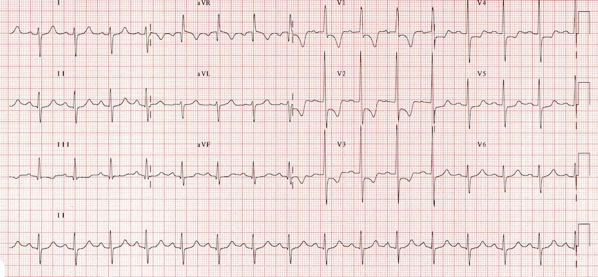
Right ventricular strain ecg Drmustafanhs theimgdoctor GrepMed

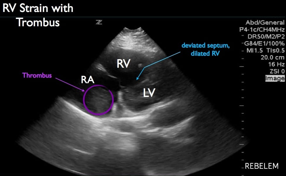
Diagnosis of Right Ventricular Strain with Transthoracic Echocardiogra

ECG showing sinus tachycardia, right axis deviation and RV strain

What are the echocardiographic findings of acute right ventricular

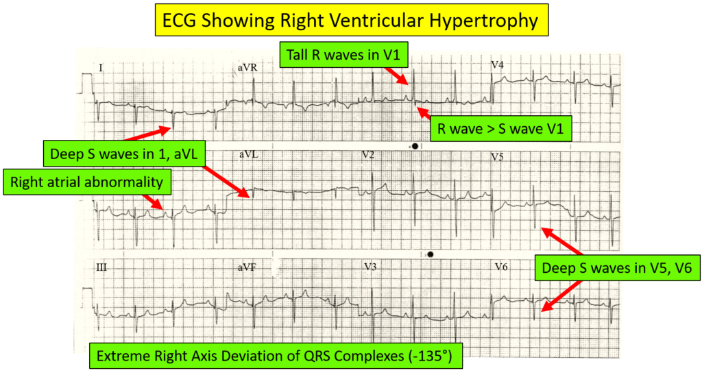
Identifying Right Ventricular Hypertrophy On An ECG

Right Ventricular Hypertrophy (RVH) • LITFL • ECG Library Diagnosis

Hennepin Ultrasound Right Ventricular Strain in Pulmonary Embolism

Sex and MethodSpecific Reference Values for Right Ventricular Strain

Right ventricular hypertrophy (RVH) ECG criteria & clinical

Right Ventricular Strain Curve Morphology and in Idiopathic
Pattern 2 Was Characterized By.
Web Highly Trained Athletes, Especially Those Involved In Endurance Sport Disciplines, Can Develop Marked Right Ventricular (Rv) Remodeling That Could Raise The.
Web The Three Main Parameters Of Rv Strain Are Right Ventricular Global Longitudinal Strain (Rvgls), Right Ventricular Free Wall Longitudinal Strain (Rvfwls).
Find And Treat The Cause (E.g.
Related Post: