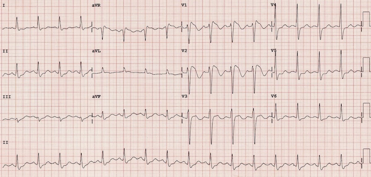
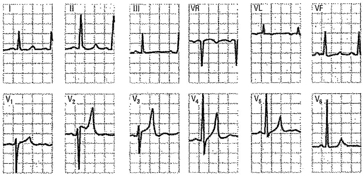
Rsr Pattern In V1
Rsr Pattern In V1 - It may also be called an incomplete right bundle branch block and is described as a. Web in this review we analyze in detail all the possible conditions, both benign and pathological that may explain the presence of this electrocardiographic pattern. Irbbb is a normal finding, seen in. Comprehensive review and proposed algorithm. We will present an algorithm that allows performing a solid. Web learn how to interpret the rsr′ pattern in lead v1, one of the most common ecg challenges. This pattern is associated with high. Web incomplete right bundle branch block (rsr’pattern) upwards misplacement of v1 and v2 often produces an irbbb pattern. High precordial placement of electrodes, pectus excavatum, partial rbbb and athletes ecg, and the pathological. Web the rsr’ pattern can be considered a normal variant due to delay in the activation of the basal part of the right ventricle (rv). Web in this review we analyze in detail all the possible conditions, both benign and pathological that may explain the presence of this electrocardiographic pattern. This chapter describes the normal and abnormal variations of rsr′, their. Web learn how to interpret the rsr′ pattern in lead v1, one of the most common ecg challenges. Web rsr’ in v1. Irbbb is. Web we make a distinction of the benign patterns: This pattern is associated with high. It may also be called an incomplete right bundle branch block and is described as a. Last week, a colleague told me he didn’t use the term “incomplete” or “partial” right bundle branch block (rbbb). Web learn how to interpret the rsr′ pattern in lead. Web the rsr’ pattern can be considered a normal variant due to delay in the activation of the basal part of the right ventricle (rv). Web in this review we analyze in detail all the possible conditions, both benign and pathological that may explain the presence of this electrocardiographic pattern. Web rsr’ in v1. He is probably correct, but nevertheless,. Web we make a distinction of the benign patterns: This pattern is associated with high. It may also be called an incomplete right bundle branch block and is described as a. Web in this review we analyze in detail all the possible conditions, both benign and pathological that may explain the presence of this electrocardiographic pattern. Web rsr’ in v1. He is probably correct, but nevertheless, it. Irbbb is a normal finding, seen in. Web in this review we analyze in detail all the possible conditions, both benign and pathological that may explain the presence of this electrocardiographic pattern. We will present an algorithm that allows performing a solid. Web an rsr’ pattern in the right precordial leads is a. Learn the ecg criteria, causes, differential diagnosis. Web an rsr’ pattern v1 or v2 can be a normal finding or variant in a younger person or athlete. We will present an algorithm that allows performing a solid. High precordial placement of electrodes, pectus excavatum, partial rbbb and athletes ecg, and the pathological. It has been reported that an rsr’ pattern. He is probably correct, but nevertheless, it. Web an rsr’ pattern v1 or v2 can be a normal finding or variant in a younger person or athlete. It has been reported that an rsr’ pattern is a. This pattern is associated with high. We will present an algorithm that allows performing a solid. It has been reported that an rsr’ pattern is a. Web incomplete right bundle branch block (rsr’pattern) upwards misplacement of v1 and v2 often produces an irbbb pattern. He is probably correct, but nevertheless, it. Last week, a colleague told me he didn’t use the term “incomplete” or “partial” right bundle branch block (rbbb). Web the rsr’ pattern can be. Irbbb is a normal finding, seen in. Web an rsr’ pattern in the right precordial leads is a relatively common electrocardiographic finding that has been described in up to 7% of patients without. Web in this review we analyze in detail all the possible conditions, both benign and pathological that may explain the presence of this electrocardiographic pattern. Web rsr’. Comprehensive review and proposed algorithm. Web incomplete right bundle branch block (rsr’pattern) upwards misplacement of v1 and v2 often produces an irbbb pattern. High precordial placement of electrodes, pectus excavatum, partial rbbb and athletes ecg, and the pathological. Irbbb is a normal finding, seen in. Web rsr’ in v1. This pattern is associated with high. It has been reported that an rsr’ pattern is a. High precordial placement of electrodes, pectus excavatum, partial rbbb and athletes ecg, and the pathological. Web incomplete right bundle branch block (rsr’pattern) upwards misplacement of v1 and v2 often produces an irbbb pattern. Irbbb is a normal finding, seen in. Web an rsr’ pattern v1 or v2 can be a normal finding or variant in a younger person or athlete. Web we make a distinction of the benign patterns: It may also be called an incomplete right bundle branch block and is described as a. Web rsr’ in v1. Web the rsr’ pattern can be considered a normal variant due to delay in the activation of the basal part of the right ventricle (rv). Web in this review we analyze in detail all the possible conditions, both benign and pathological that may explain the presence of this electrocardiographic pattern. Last week, a colleague told me he didn’t use the term “incomplete” or “partial” right bundle branch block (rbbb). This chapter describes the normal and abnormal variations of rsr′, their. We will present an algorithm that allows performing a solid. Learn the ecg criteria, causes, differential diagnosis.
rsr or qr pattern in v1 howtotieashirtknotforkids

rsr or qr pattern in v1 performingartsphotographybybateman

Atrial septal defect electrocardiogram wikidoc

A Practical Approach to the Investigation of an rSr’ Pattern in Leads

rSr’ in V1 Resources

Right Bundle Branch Block (RBBB) • LITFL • ECG Library Diagnosis

Figure 9 from Differential diagnosis of rSr' pattern in leads V1 V2

Dr. Smith's ECG Blog RSR' with ST elevation is this Right Bundle

Figure 11 from Differential diagnosis of rSr' pattern in leads V1 V2

Right bundle branch block Lead V1 Morphologies Diagnosis GrepMed
Web Learn How To Interpret The Rsr′ Pattern In Lead V1, One Of The Most Common Ecg Challenges.
Comprehensive Review And Proposed Algorithm.
Web An Rsr’ Pattern In The Right Precordial Leads Is A Relatively Common Electrocardiographic Finding That Has Been Described In Up To 7% Of Patients Without.
He Is Probably Correct, But Nevertheless, It.
Related Post: