Ribosomes Drawing
Ribosomes Drawing - They are sites of protein synthesis. Web what are ribosomes in biology, where are they found & what do they do: Web describe the different steps in protein synthesis. The colored balls at the top of this diagram represent different amino acids. These clusters, called polysomes, are held together by messenger rna (mrna). Discuss the role of ribosomes in protein synthesis. Web describe the different steps in protein synthesis. Discuss the role of ribosomes in protein synthesis. Web ribosomes | structure of ribosomes | easy step by step diagram of ribosomes | class 9th | biology. Web shape, size and function. Web shape, size and function. The synthesis of proteins consumes more of a cell’s energy than any other metabolic process. Long chains of amino acids fold and function as proteins in cells. Discuss the role of ribosomes in protein synthesis. Ribosomes are roughly spherical with a diameter of ~20 nm, they can be seen only with the electron microscope. Discuss the role of ribosomes in protein synthesis. Ribosomes are composed of special proteins and nucleic acids. During translation, the two subunits come together around a mrna molecule, forming a complete ribosome. The small and large ribosomal subunits. Ribosomes consist of two major components: Types of ribosomes based on locations. Describe the different steps in protein synthesis. It comprises of two sections, known as subunits. 1 is an electron micrograph showing clusters of ribosomes. Cutaway drawing of a eukaryotic plant cell. This chain of amino acids then folds to form a complex 3d structure. They are structures containing approximately equal amounts of rna and proteins and serve as a scaffold for the ordered interaction of the numerous. They are sites of protein synthesis. Cutaway drawing of a eukaryotic plant cell. The synthesis of proteins consumes more of a cell’s energy than. Web ribosomes are made of proteins and ribonucleic acid (abbreviated as rna), in almost equal amounts. 35k views 5 years ago. The colored balls at the top of this diagram represent different amino acids. Discuss the role of ribosomes in protein synthesis. Pattern of a cell endoplasmic reticulum. The ribosome moves forward on the mrna, codon by codon, as it is read and translated into a polypeptide (protein chain). 35k views 5 years ago. Learn more about ribosomes and how they work, see the khan academy. A large and a small subunit. Ribosomes consist of two major components: Web browse 17 ribosome drawing photos and images available, or start a new search to explore more photos and images. The ribosome moves forward on the mrna, codon by codon, as it is read and translated into a polypeptide (protein chain). This chain of amino acids then folds to form a complex 3d structure. Ribosomes occur both as free particles. Web ribosomes are made of proteins and ribonucleic acid (abbreviated as rna), in almost equal amounts. A ribosome is a complex cellular mechanism used to translate genetic code into chains of amino acids. 35k views 5 years ago. The colored balls at the top of this diagram represent different amino acids. Lady of hats from wikipedia; They are sites of protein synthesis. Discuss the role of ribosomes in protein synthesis. Web describe the different steps in protein synthesis. Web check out this book to learn how the structure of the ribosome was discovered; During translation, the two subunits come together around a mrna molecule, forming a complete ribosome. Types of ribosomes based on locations. The synthesis of proteins consumes more of a cell’s energy than any other metabolic process. Pattern of a cell endoplasmic reticulum. The ribosome moves forward on the mrna, codon by codon, as it is read and translated into a polypeptide (protein chain). In this video, you will learn how to draw. Biology for science majors i. How to draw ribosomes how to draw cell • how to draw animal cell.more. Web browse 17 ribosome drawing photos and images available, or start a new search to explore more photos and images. Web ribosomes | structure of ribosomes | easy step by step diagram of ribosomes | class 9th | biology. The synthesis of proteins consumes more of a cell’s energy than any other. Web click to view a research level microscope image, interpreted using cimr gridpoint technology. Ribosomes occur both as free particles in prokaryotic and eukaryotic cells and as particles attached to the membranes of the endoplasmic reticulum in eukaryotic cells. Web describe the different steps in protein synthesis. Web describe the different steps in protein synthesis. Learn more about ribosomes and how they work, see the khan academy. It's an educational video from 9th biology ptb. Ribosomes are roughly spherical with a diameter of ~20 nm, they can be seen only with the electron microscope. The synthesis of proteins consumes more of a cell’s energy than any other metabolic process. Discuss the role of ribosomes in protein synthesis. Lady of hats from wikipedia; The small and large ribosomal subunits.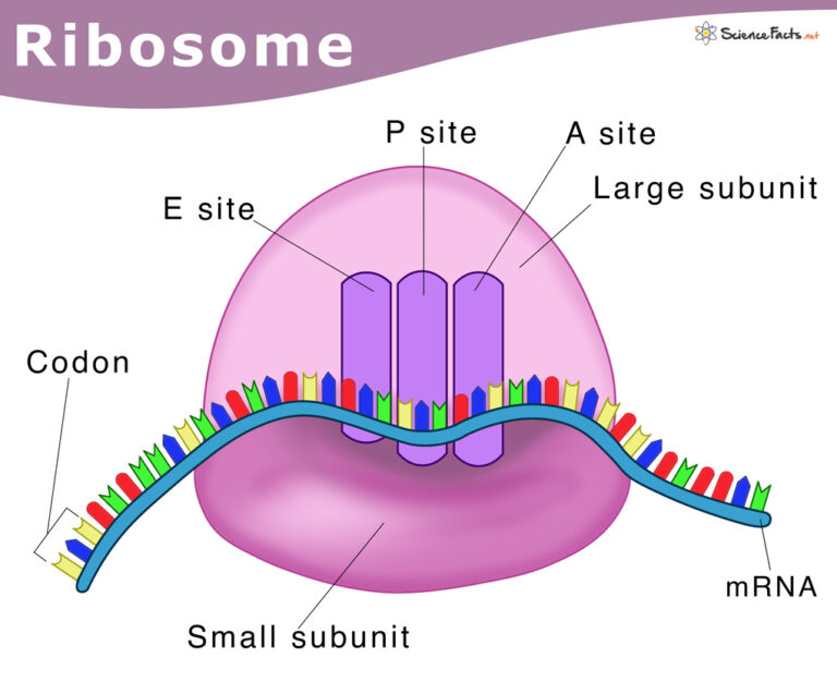
Ribosomes Definition, Structure, & Functions, with Diagram

The structure of the ribosome Infographics on Vector Image
Biology. Drawing of ribosome / Biología. Ribosoma
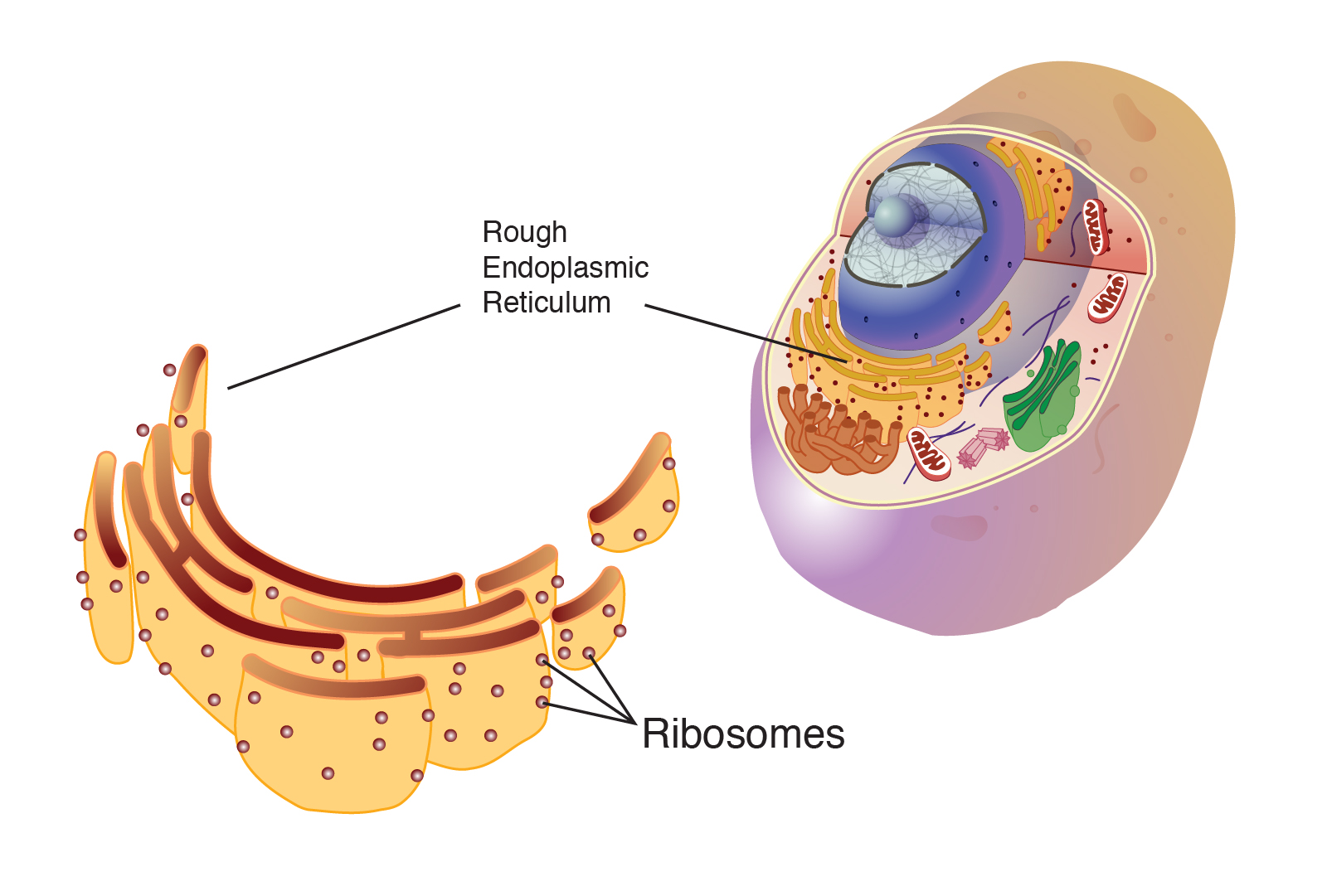
Ribosome Talking Glossary of Terms NHGRI
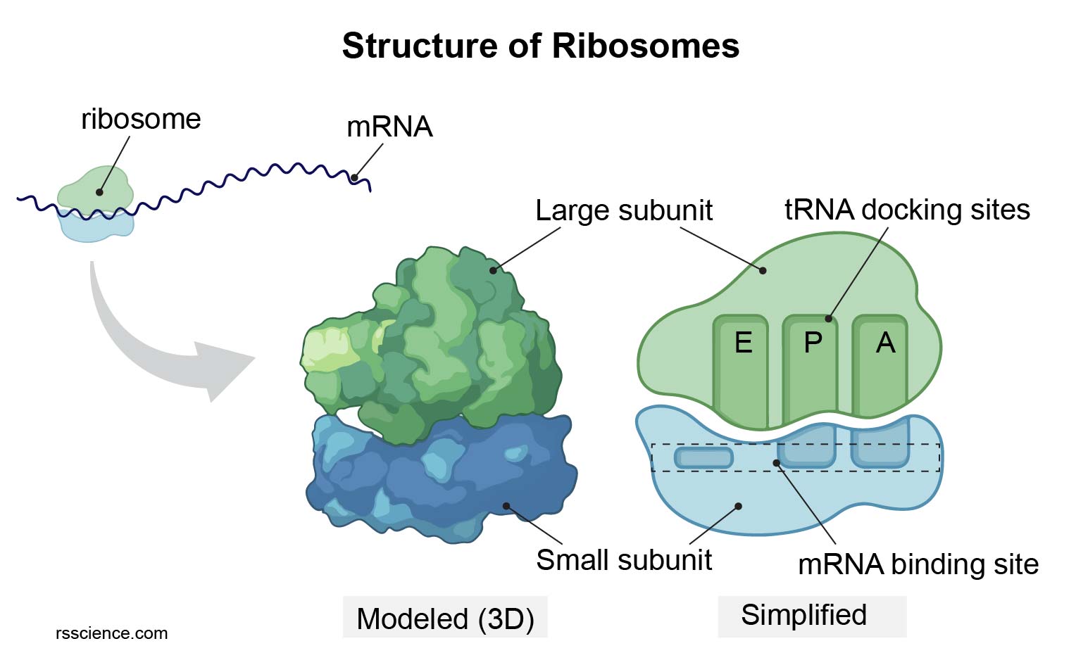
Ribosome protein factory definition, function, structure and biology

The structure of the ribosome Infographics Vector Image
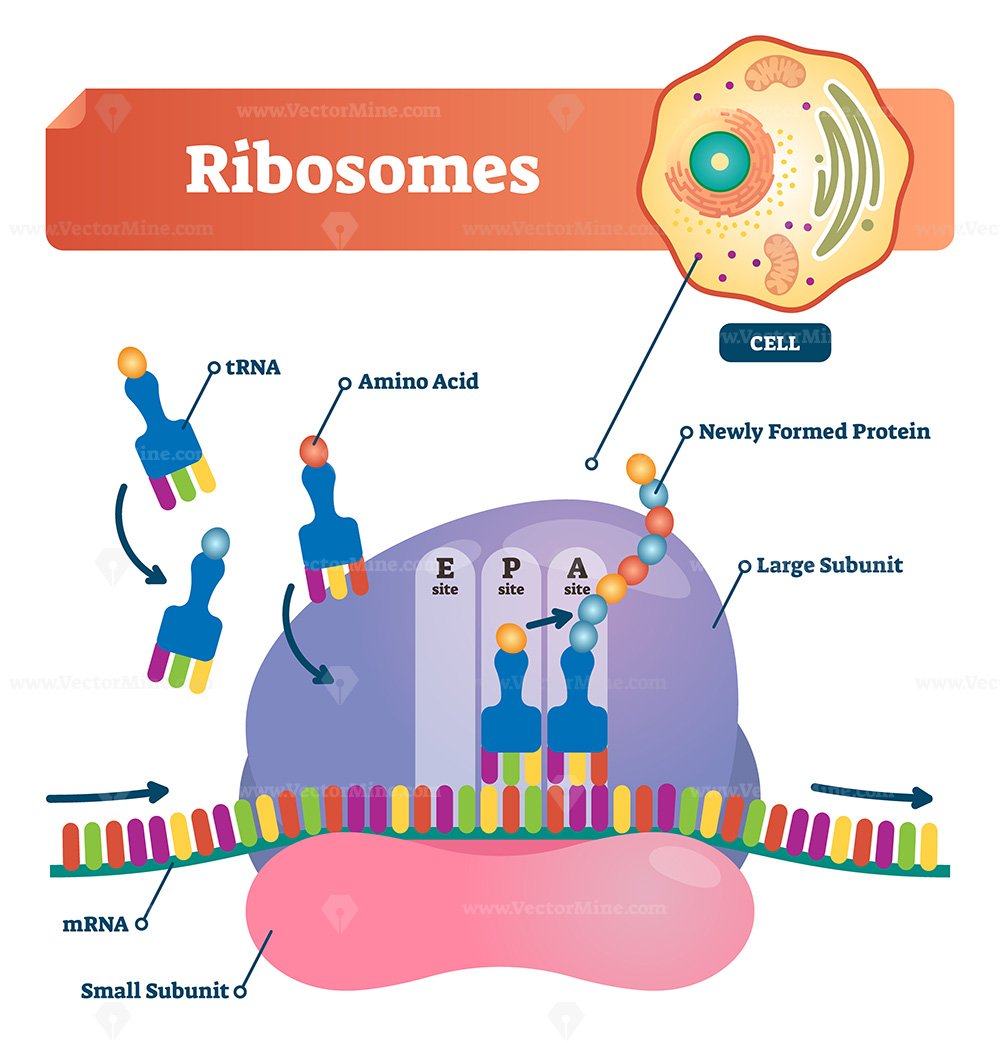
Ribosomes vector illustration VectorMine
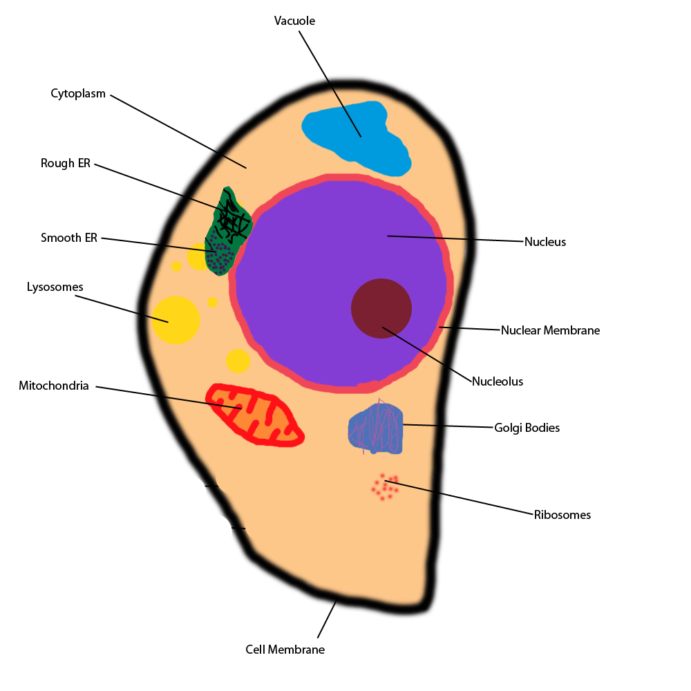
Ribosome Diagram ClipArt Best

Charlie Garton Science 8 Ribosomes & Endoplasmic Reticulum
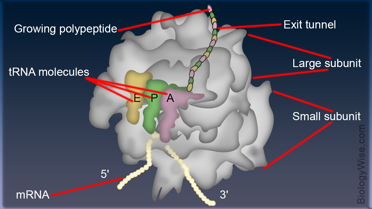
Structure of Ribosome Biology Wise
Pattern Of A Cell Endoplasmic Reticulum.
They Are Sites Of Protein Synthesis.
The Tinier Subunit Is The Place The Mrna Binds And It Decodes, Whereas The Bigger Subunit Is The Place The Amino Acids Are Included.
During Translation, The Two Subunits Come Together Around A Mrna Molecule, Forming A Complete Ribosome.
Related Post: