Reticulonodular Pattern Chest X Ray
Reticulonodular Pattern Chest X Ray - Web an explanation of alveolar vs. Part of an underlying reticulonodular pattern. Web chest radiographs show nonspecific bilateral reticular, reticulonodular, or alveolar opacities that may be distributed diffusely or have a lower lung predominance. What is an interstitial lung pattern? Web nodular opacification is one of the broad patterns of pulmonary opacification that can be described on a chest radiograph or chest ct. Nodular opacification in the lung may be a. Web infection of the lower respiratory tract, acquired by way of the airways and confined to the lung parenchyma and airways, typically presents radiologically as one of. This may be used to describe a regional pattern or a. This finding can be asymptomatic or have severe and life threatening symptoms. Web a reticular pattern on the radiograph may result from summation of smooth or irregular linear opacities, cystic spaces, or both. Most lung nodules are benign (not cancerous). Nodular opacification in the lung may be a. Web in patients with suspected diffuse parenchymal lung disease (based on clinical findings, chest radiography, or pulmonary function abnormalities), indications for hrct of the. Web an interstitial lung pattern is a regular descriptive term used when reporting a plain chest radiograph. Web a lung (pulmonary). Nodular opacification in the lung may be a. In approximately 10% of patients with. Web a lung (pulmonary) nodule is an abnormal growth that forms in a lung. This finding can be asymptomatic or have severe and life threatening symptoms. The micronodules may either be. Web nodular opacification is one of the broad patterns of pulmonary opacification that can be described on a chest radiograph or chest ct. Ct scans of your chest also may have been done, and those should also be. This may be used to describe a regional pattern or a. You may have one nodule on the lung or several nodules.. Imaging findings are varied and nonspecific. In approximately 10% of patients with. Nodules may develop in one lung or both. Web nodular opacification is one of the broad patterns of pulmonary opacification that can be described on a chest radiograph or chest ct. Ct scans of your chest also may have been done, and those should also be. Web a lung (pulmonary) nodule is an abnormal growth that forms in a lung. Most lung nodules are benign (not cancerous). Web an explanation of alveolar vs. This finding can be asymptomatic or have severe and life threatening symptoms. Web a reticular pattern on the radiograph may result from summation of smooth or irregular linear opacities, cystic spaces, or both. Rarely, pulmonary nodules are a sign of lung cancer. Web a reticular pattern on the radiograph may result from summation of smooth or irregular linear opacities, cystic spaces, or both. Web list and identify on a chest radiograph and computed tomographic (ct) scan the four patterns of interstitial lung disease (ild): Web histological features of cmv pneumonia consist of areas. Web histological features of cmv pneumonia consist of areas of acute interstitial pneumonia, dad and haemorrhage. In approximately 10% of patients with. Web chest radiographs show nonspecific bilateral reticular, reticulonodular, or alveolar opacities that may be distributed diffusely or have a lower lung predominance. A reticulonodular interstitial pattern is produced by either overlap of reticular shadows or by the presence. Web a reticulonodular interstitial pattern is an imaging descriptive term that can be used in thoracic radiographs or ct scans when are there is an overlap of reticular shadows with nodular shadows. The micronodules may either be. Nodules may develop in one lung or both. A reticulonodular interstitial pattern is produced by either overlap of reticular shadows or by the. Most lung nodules are benign (not cancerous). Ct scans of your chest also may have been done, and those should also be. Imaging findings are varied and nonspecific. Web list and identify on a chest radiograph and computed tomographic (ct) scan the four patterns of interstitial lung disease (ild): Web an interstitial lung pattern is a regular descriptive term used. Web a reticular pattern on the radiograph may result from summation of smooth or irregular linear opacities, cystic spaces, or both. Ct scans of your chest also may have been done, and those should also be. Web infection of the lower respiratory tract, acquired by way of the airways and confined to the lung parenchyma and airways, typically presents radiologically. Nodules may develop in one lung or both. This may be used to describe a regional pattern or a. Imaging findings are varied and nonspecific. Ct scans of your chest also may have been done, and those should also be. Web a lung (pulmonary) nodule is an abnormal growth that forms in a lung. Part of an underlying reticulonodular pattern. Web infection of the lower respiratory tract, acquired by way of the airways and confined to the lung parenchyma and airways, typically presents radiologically as one of. Web histological features of cmv pneumonia consist of areas of acute interstitial pneumonia, dad and haemorrhage. The micronodules may either be. Web list and identify on a chest radiograph and computed tomographic (ct) scan the four patterns of interstitial lung disease (ild): Web nodular opacification is one of the broad patterns of pulmonary opacification that can be described on a chest radiograph or chest ct. Web chest radiographs show nonspecific bilateral reticular, reticulonodular, or alveolar opacities that may be distributed diffusely or have a lower lung predominance. Web an explanation of alveolar vs. Rarely, pulmonary nodules are a sign of lung cancer. In approximately 10% of patients with. Web an interstitial lung pattern is a regular descriptive term used when reporting a plain chest radiograph.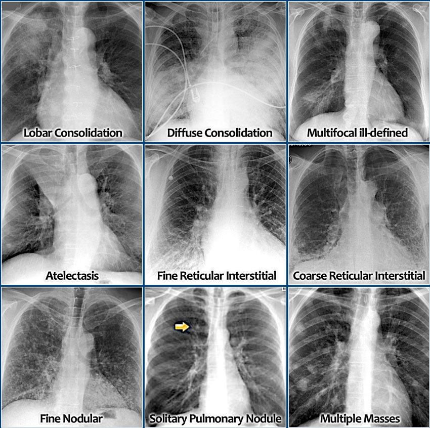
The Radiology Assistant Chest XRay Lung disease
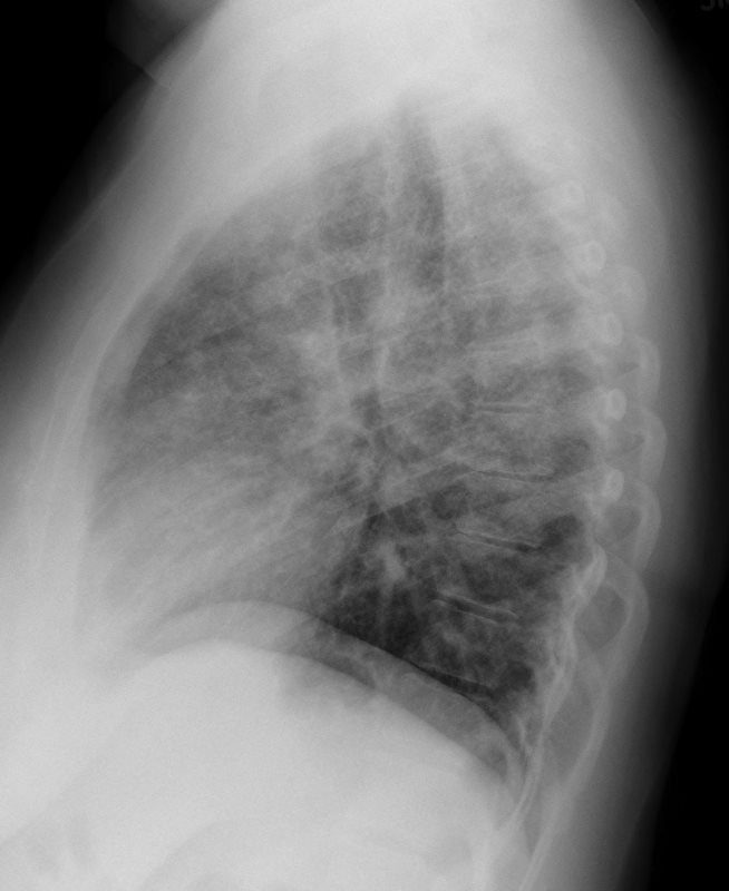
CXR Reticulonodular Pattern Lungs

Chest Xray showing bilateral reticulonodular opacities with some

Chest radiograph demonstrated diffuse infiltrates with reticular and

Chest Xrays reticulonodular pattern with perihilar distribution
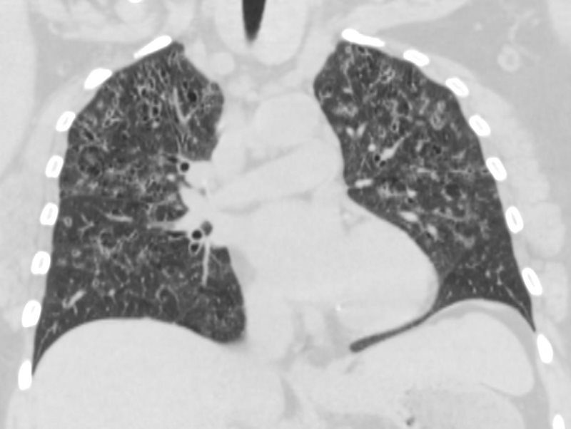
CXR Reticulonodular Pattern Lungs
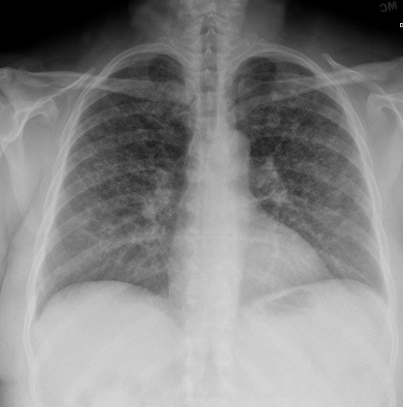
CXR Reticulonodular Pattern Lungs
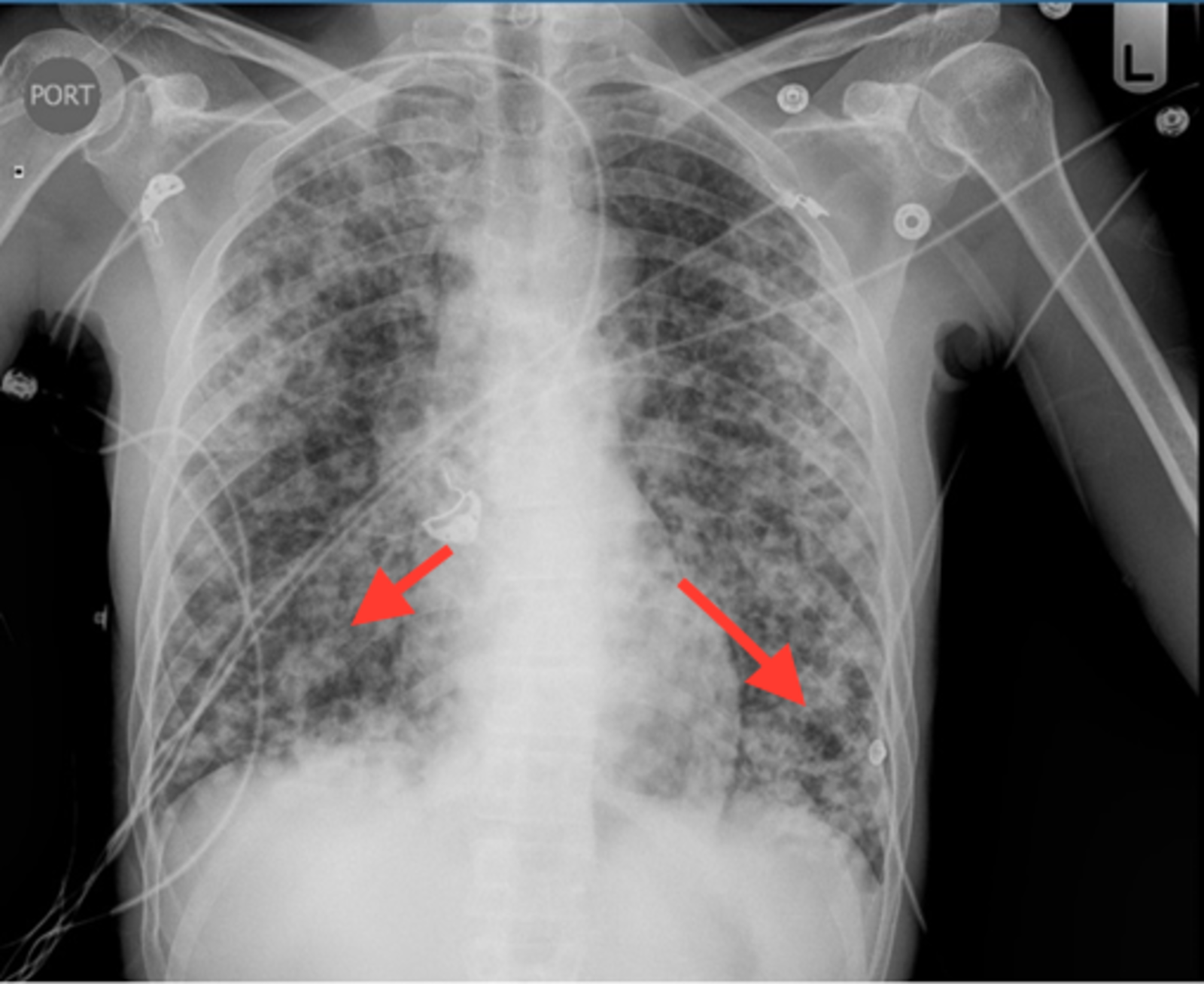
Cureus Recurrent Pneumocystis Pneumonia with Radiographic

a) CXR in anteroposterior view shows bilateral reticular pattern with
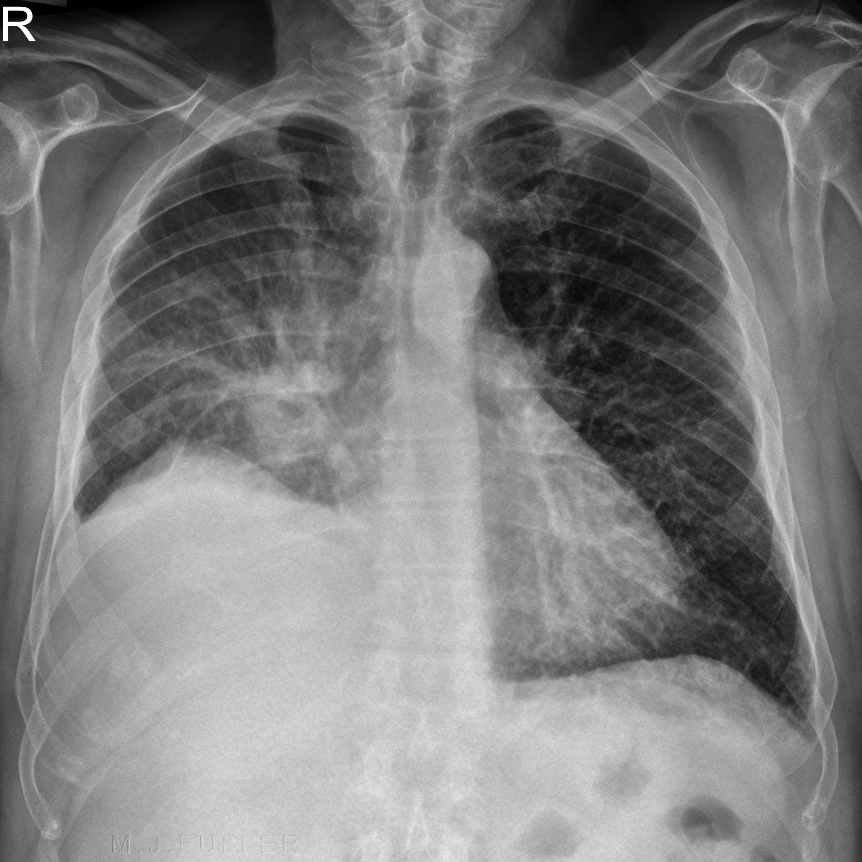
Interstitial vs Alveolar Lung Patterns wikiRadiography
Web A Reticular Pattern On The Radiograph May Result From Summation Of Smooth Or Irregular Linear Opacities, Cystic Spaces, Or Both.
The Others, Linear Opacification And Airway Opacification Are Discussed Separately.
Most Lung Nodules Are Benign (Not Cancerous).
Nodular Opacification In The Lung May Be A.
Related Post: