Pulmonary Disease Pattern On Ecg
Pulmonary Disease Pattern On Ecg - Web patients with chronic obstructive pulmonary disease (copd) often have abnormal ecgs. A pulmonary embolism (pe) is a blood clot in. Web a better understanding of the ecg changes in copd may improve interpretation of ecg in these patients and help revealing the dominant pathophysiology. Web the electrocardiogram is often abnormal in patients who have chronic obstructive pulmonary disease. Signs & symptomsfinding supportreal storiespah community Ecg changes commonly associated with pulmonary diseases such as copd. Our aim was to separate the effects on ecg by airway obstruction,. Web electrocardiographic (ecg) abnormalities associated with chronic obstructive pulmonary disease (copd) include right atrial enlargement, right. Web specific electrocardiographic abnormalities and cardiac arrhythmias are prevalent in chronic obstructive pulmonary disease. Web ecg abnormalities are common in patients with pulmonary embolism, with the most frequent being sinus tachycardia, right ventricular strain, and the classic s1q3t3. Web specific electrocardiographic abnormalities and cardiac arrhythmias are prevalent in chronic obstructive pulmonary disease. Web copd can cause electrocardiographic changes due to factors including lung hyperinflation. The underlying pathophysiology is complex. Signs & symptomsfinding supportreal storiespah community Web what an ecg can tell you about pulmonary embolism. Web electrocardiographic (ecg) abnormalities associated with chronic obstructive pulmonary disease (copd) include right atrial enlargement, right. Web pulmonary embolism (submassive or massive) may cause acute right ventricle overload or failure, which manifests classically (but not commonly) as right axis deviation (r > s in. Web the electrocardiogram is often abnormal in patients who have chronic obstructive pulmonary disease. Ecg findings. Web electrocardiographic (ecg) abnormalities associated with chronic obstructive pulmonary disease (copd) include right atrial enlargement, right. A pulmonary embolism (pe) is a blood clot in. Web what an ecg can tell you about pulmonary embolism. Ecg changes commonly associated with pulmonary diseases such as copd. Web pulmonary embolism (submassive or massive) may cause acute right ventricle overload or failure, which. Signs & symptomsfinding supportreal storiespah community Web (1) airway obstruction by fev% (percentage of forced expiratory volume)_predicted, (2) emphysema by residual volume/total lung capacity and residual volume (percent of. Web the following ecg signs reflecting ccp were collected: Web copd can cause electrocardiographic changes due to factors including lung hyperinflation. Web what an ecg can tell you about pulmonary embolism. Web copd can cause electrocardiographic changes due to factors including lung hyperinflation. Web electrocardiographic (ecg) abnormalities associated with chronic obstructive pulmonary disease (copd) include right atrial enlargement, right. Web ecg abnormalities are common in patients with pulmonary embolism, with the most frequent being sinus tachycardia, right ventricular strain, and the classic s1q3t3. Web patients with chronic obstructive pulmonary disease (copd). These changes can be present on the electrocardiograms of patients. Web pulmonary embolism (submassive or massive) may cause acute right ventricle overload or failure, which manifests classically (but not commonly) as right axis deviation (r > s in. Copd particularly chronic bronchitis was the commonest respiratory. A pulmonary embolism (pe) is a blood clot in. Web sinus tachycardia is the. Copd particularly chronic bronchitis was the commonest respiratory. Web the following ecg signs reflecting ccp were collected: Our aim was to separate the effects on ecg by airway obstruction,. Web patients with chronic obstructive pulmonary disease (copd) often have abnormal ecgs. Web copd can cause electrocardiographic changes due to factors including lung hyperinflation. The underlying pathophysiology is complex. Copd particularly chronic bronchitis was the commonest respiratory. These changes can be present on the electrocardiograms of patients. Web pulmonary embolism (submassive or massive) may cause acute right ventricle overload or failure, which manifests classically (but not commonly) as right axis deviation (r > s in. Web patients with chronic obstructive pulmonary disease (copd) often. Web (1) airway obstruction by fev% (percentage of forced expiratory volume)_predicted, (2) emphysema by residual volume/total lung capacity and residual volume (percent of. Web what an ecg can tell you about pulmonary embolism. Our aim was to separate the effects on ecg by airway obstruction,. Web a better understanding of the ecg changes in copd may improve interpretation of ecg. Web ecg abnormalities are common in patients with pulmonary embolism, with the most frequent being sinus tachycardia, right ventricular strain, and the classic s1q3t3. A pulmonary embolism (pe) is a blood clot in. Web what an ecg can tell you about pulmonary embolism. Web sinus tachycardia is the most common ecg finding in pulmonary embolism. Web the following ecg signs. Web pulmonary embolism (submassive or massive) may cause acute right ventricle overload or failure, which manifests classically (but not commonly) as right axis deviation (r > s in. Web sinus tachycardia is the most common ecg finding in pulmonary embolism. Copd particularly chronic bronchitis was the commonest respiratory. Web the electrocardiogram is often abnormal in patients who have chronic obstructive pulmonary disease. Web patients with chronic obstructive pulmonary disease (copd) often have abnormal ecgs. A pulmonary embolism (pe) is a blood clot in. Web ecg abnormalities are common in patients with pulmonary embolism, with the most frequent being sinus tachycardia, right ventricular strain, and the classic s1q3t3. Web electrocardiographic (ecg) abnormalities associated with chronic obstructive pulmonary disease (copd) include right atrial enlargement, right. Web the following ecg signs reflecting ccp were collected: The underlying pathophysiology is complex. Web (1) airway obstruction by fev% (percentage of forced expiratory volume)_predicted, (2) emphysema by residual volume/total lung capacity and residual volume (percent of. These changes can be present on the electrocardiograms of patients. Web a better understanding of the ecg changes in copd may improve interpretation of ecg in these patients and help revealing the dominant pathophysiology. Web specific electrocardiographic abnormalities and cardiac arrhythmias are prevalent in chronic obstructive pulmonary disease. Web sinus tachycardia is the most common ecg finding in pulmonary embolism. Web copd can cause electrocardiographic changes due to factors including lung hyperinflation.
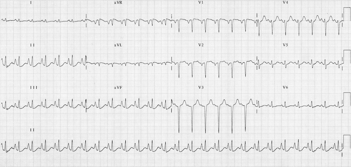
ECG in Chronic Obstructive Pulmonary Disease • LITFL • ECG Library

pulmonary disease pattern ecg Hình ảnh có liên quan Diseases Club

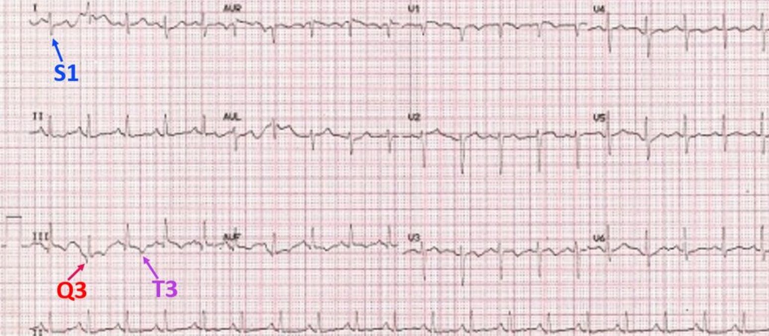
S1Q3T3 EKG Classic Pattern in Pulmonary Embolism (Example). Pulmonary

Pulmonary embolism and S1Q3 pattern Cardiocases

ECG Changes in Pulmonary Embolism New Health Advisor

ECG in Chronic Obstructive Pulmonary Disease • LITFL • ECG Library

Pulmonary Embolism (PE) Causes, symptoms, diagnosis, treatment

S1Q3T3 on ECG in a patient with Acute Pulmonary Embolism GrepMed

Dr. Smith's ECG Blog Acute Pulmonary Edema, Respiratory Failure, and LBBB

The ECG's of Pulmonary Embolism Resus
Web What An Ecg Can Tell You About Pulmonary Embolism.
Signs & Symptomsfinding Supportreal Storiespah Community
Ecg Changes Commonly Associated With Pulmonary Diseases Such As Copd.
Web Pulmonary Embolism (Submassive Or Massive) May Cause Acute Right Ventricle Overload Or Failure, Which Manifests Classically (But Not Commonly) As Right Axis Deviation (R > S In.
Related Post: