Protein Molecule Drawing
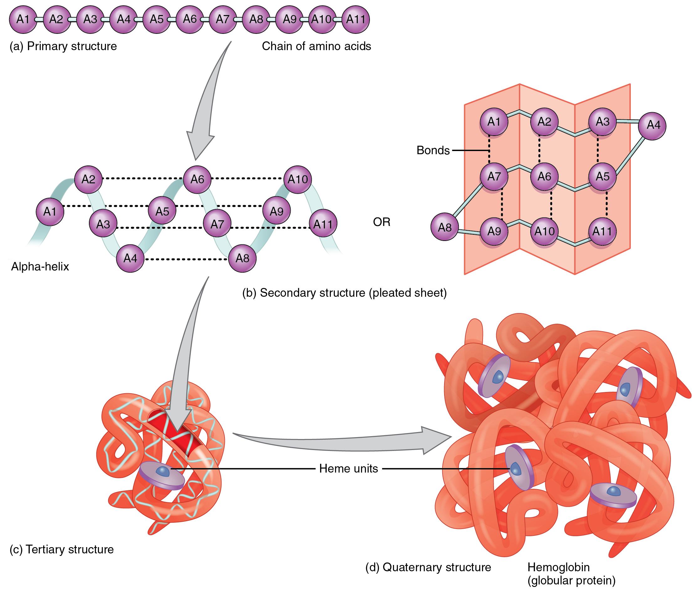
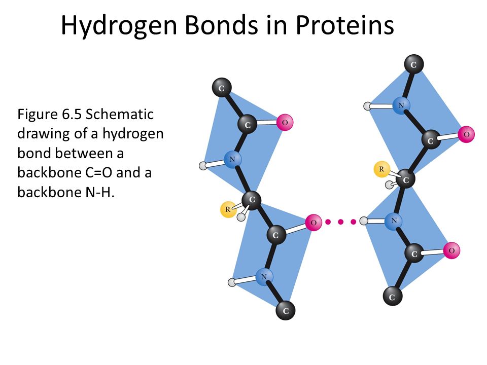
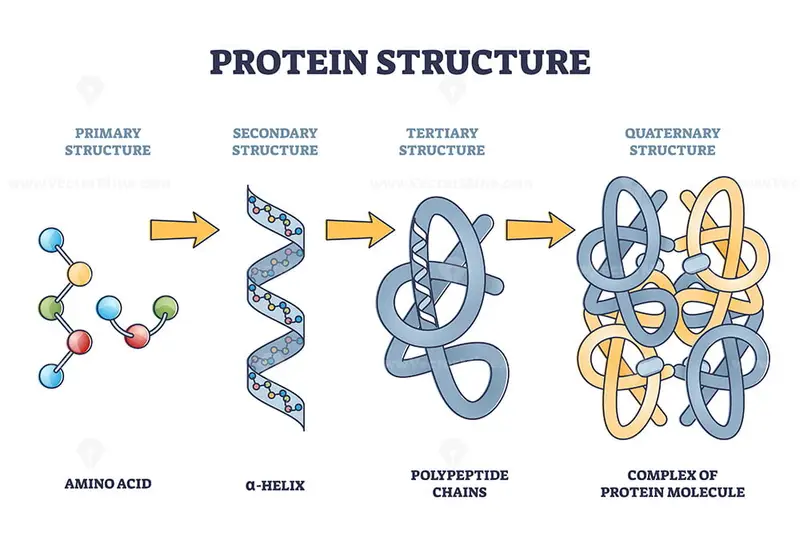
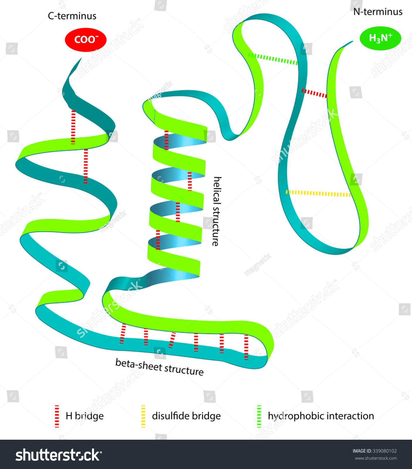
Protein Molecule Drawing - Web molview is a really nice tool that allows you to draw and visualize molecules in both 2d and 3d; Depending on what they are interested in looking at they will pick different ways to draw and display the protein. Web explore and visualize protein structures using protein imager web app. Web the primary structure of proteins. Web deep learning methods excel at predicting molecular structures with high efficiency. It folds up into a coil, and the shape of that coil influences its biological activity. Sometimes a biochemist may be interested in how the chain of amino acids coils up. You have two ways to. Primary, secondary, tertiary, and quaternary. Web search by structure or substructure. Primary, secondary, tertiary, and quaternary. Proteins, nucleic acids, and lipid membranes are shown; Web scientists have many different ways to draw and look at protein shapes and structure. Navigate through molecular structures, analyze interactions, and gain insights into protein functionality. For example, alphafold predicts protein structures with atomic accuracy 1, enabling new structural biology. Goodsell integrate information from structural biology, microscopy and biophysics to simulate detailed views of the molecular structure of living cells. Web interactive molecular modeling system for analysis and presentation graphics of molecular structures and related data, including densitymaps, sequence alignments, trajectories, and docking results; Web explore and visualize protein structures using protein imager web app. The resulting smiles or inchi. It folds up into a coil, and the shape of that coil influences its biological activity. Proteins, nucleic acids, and lipid membranes are shown; The resulting smiles or inchi string may be used to search for matching molecules in the pdb chemical component dictionary. Depending on what they are interested in looking at they will pick different ways to draw. Web as we mentioned in the last article on proteins and amino acids, the shape of a protein is very important to its function. Depending on what they are interested in looking at they will pick different ways to draw and display the protein. Web the building blocks of proteins are amino acids, which are small organic molecules that consist. Once you’ve drawn a molecule, you can click the 2d to 3d button to convert the molecule into a 3d model which is then displayed in the viewer. Primary, secondary, tertiary, and quaternary. Depending on what they are interested in looking at they will pick different ways to draw and display the protein. Web explore and visualize protein structures using. Web explore and visualize protein structures using protein imager web app. Web protein folding and structure. These illustrations are free for use under. Free for noncommercial use, available for windows, linux, and mac os x. Web search by structure or substructure. Each type of protein has a unique sequence of amino acids, exactly the same from one molecule to the next. Web the watercolor paintings of david s. Vast identifies 3d domains (substructures) within each protein structure in the molecular modeling database (mmdb), and then finds other protein structures that have one or more similar 3d domains, using purely geometric criteria.. To understand how a protein gets its final shape or conformation, we need to understand the four levels of protein structure: Each type of protein has a unique sequence of amino acids, exactly the same from one molecule to the next. Navigate through molecular structures, analyze interactions, and gain insights into protein functionality. Web protein folding and structure. For example,. These illustrations are free for use under. Sometimes a biochemist may be interested in how the chain of amino acids coils up. Each type of protein has a unique sequence of amino acids, exactly the same from one molecule to the next. Web explore and visualize protein structures using protein imager web app. Vast identifies 3d domains (substructures) within each. Web when i generate protein schematics for figures, i emphasize the main points of protein structures without getting bogged down in the details. For a short (4 minutes) introduction video. Web use the chemical sketch tool to draw or edit a molecule. Web interactive molecular modeling system for analysis and presentation graphics of molecular structures and related data, including densitymaps,. Can also be used for visualizing macromolecules; Whereas earlier versions of the company’s software could model how the strands of amino acids making up a protein fold into its final 3d shape, the new version reveals how folded. Primary, secondary, tertiary, and quaternary. I try to depict the overall shape of the molecule by drawing the structures as simple shapes. Once you’ve drawn a molecule, you can click the 2d to 3d button to convert the molecule into a 3d model which is then displayed in the viewer. The tools tab allows you to see calculated properties and spectroscopy data However, for drawing the structures of proteins, we usually twist it. Vast identifies 3d domains (substructures) within each protein structure in the molecular modeling database (mmdb), and then finds other protein structures that have one or more similar 3d domains, using purely geometric criteria. Web explore and visualize protein structures using protein imager web app. For example, alphafold predicts protein structures with atomic accuracy 1, enabling new structural biology. You have two ways to. Primary, secondary, tertiary, and quaternary. Protein viewer is based on biojs. Web proteins are among the most abundant organic molecules in living systems and are way more diverse in structure and function than other classes of macromolecules. Goodsell integrate information from structural biology, microscopy and biophysics to simulate detailed views of the molecular structure of living cells. The resulting smiles or inchi string may be used to search for matching molecules in the pdb chemical component dictionary.
2.23 Protein Structure Nutrition

Proteins Drawing at GetDrawings Free download

Protein Molecule Structure

Protein structure levels from amino acid to complex molecule outline

Protein structure vector illustration Protein Muffins, Protein Snacks

The Basics of Protein Structure and Function Interactive Biology
Best Protein Molecule Illustrations, RoyaltyFree Vector Graphics

Protein Illustrations and Visualization Ask A Biologist

Protein Atomic Structure

Illustrated Structure Of A Protein Molecule Photo libre de droits
Web Molview Is A Really Nice Tool That Allows You To Draw And Visualize Molecules In Both 2D And 3D;
Web This Page Explains How Amino Acids Combine To Make Proteins And What Is Meant By The Primary, Secondary, Tertiary And Quaternary Structures Of Proteins.
Each Type Of Protein Has A Unique Sequence Of Amino Acids, Exactly The Same From One Molecule To The Next.
A Single Cell Can Contain Thousands Of Proteins, Each With A Unique Function.
Related Post:
