Phospholipid Bilayer Drawing
Phospholipid Bilayer Drawing - The head is a phosphate molecule that is attracted to water ( hydrophilic ). Role of the phospholipid bilayer in the cell membrane function. How easily these molecules can cross the membrane depends on their size and polarity. Web image of the plasma membrane, showing the phospholipid bilayer with peripheral and integral membrane proteins, glycoproteins (proteins with a carbohydrate attached), glycolipids (lipids with a carbohydrate attached), and cholesterol molecules. In eukaryotic cells, the plasma membrane surrounds a cytoplasm filled with ribosomes and organelles. So, what determines what can go in or out? Can anything or everything enter or leave? The 3 proteins have lines with the label integral membrane proteins. Their hydrophobic tails facing inwards and hydrophilic heads outwards. Web the phospholipid bilayer is the fundamental structure of the plasma membrane. The plasma membrane is the border between the interior and exterior of a cell. The two layers of phospholipids are loosely held together by weak hydrophobic interactions between the hydrocarbon tails allowing some membrane fluidity. Web the lipid bilayer (or phospholipid bilayer) is a thin polar membrane made of two layers of lipid molecules. Web a phospholipid bilayer is composed. Web a phospholipid bilayer is composed of two layers of phospholipids; Web the main component (building block) of cell membranes. There are two important parts of a phospholipid: Learn vocabulary, terms, and more with flashcards, games, and other study tools. These membranes are flat sheets that form a continuous barrier around all cells. Click the card to flip 👆. Web what are phospholipids? Structure of the cell membrane. Web the molecular details of phospholipids and their variations can be drawn as shown. The head is a phosphate molecule that is attracted to water ( hydrophilic ). Web image of the plasma membrane, showing the phospholipid bilayer with peripheral and integral membrane proteins, glycoproteins (proteins with a carbohydrate attached), glycolipids (lipids with a carbohydrate attached), and cholesterol molecules. Click the card to flip 👆. The phospholipid bilayer with hydrophobic 'tails' and hydrophilic 'heads' of the phospholipid.more. One of the proteins is shown with a channel in it.. Their hydrophobic tails facing inwards and hydrophilic heads outwards. So, what determines what can go in or out? Click the card to flip 👆. Web image of the plasma membrane, showing the phospholipid bilayer with peripheral and integral membrane proteins, glycoproteins (proteins with a carbohydrate attached), glycolipids (lipids with a carbohydrate attached), and cholesterol molecules. Learn vocabulary, terms, and more. Image modified from openstax biology. Organelles are structures that are themselves encased in membranes. All cells have a plasma membrane. So, what determines what can. The head is a phosphate molecule that is attracted to water ( hydrophilic ). The plasma membrane is made up primarily of a bilayer of phospholipids with embedded proteins, carbohydrates, glycolipids, and glycoproteins, and, in animal cells, cholesterol ( figure 6.1.1 6.1. The two layers of phospholipids are loosely held together by weak hydrophobic interactions between the hydrocarbon tails allowing some membrane fluidity. As such, it controls passage of various molecules—including sugars, amino acids,. Web a phospholipid bilayer is composed of two layers of phospholipids; Want to join the conversation? Web the lipid bilayer (or phospholipid bilayer) is a thin polar membrane made of two layers of lipid molecules. Learn vocabulary, terms, and more with flashcards, games, and other study tools. Web browse phospholipid bilayer (also referred to as the lipid bilayer) icons for. Web when drawing and labeling a diagram of the plasma membrane you should be sure to include: Can anything or everything enter or leave? Image modified from openstax biology. We will explore its components, structure, functions, examples & all about it. Web so what is its role? One of the proteins is shown with a channel in it. Web start studying label the phospholipid bilayer. Phospholipids and the phospholipid bilayer. Arc membrane (phospholipid bilayer, endocytosing) arc membrane (phospholipid bilayer, filled, landscape) arc membrane (phospholipid bilayer, filled, portrait) The hydrophilic (polar) head group and hydrophobic tails (fatty acid chains) are depicted in. Can anything or everything enter or leave? Web start studying label the phospholipid bilayer. Attached to the glycerol on one side are two fatty acids (shown in yellow at number 3). Some organelles (nuclei, mitochondria, chloroplasts) are even surrounded by double membranes. This membrane surrounds the cell. Web when drawing and labeling a diagram of the plasma membrane you should be sure to include: The hydrophilic (polar) head group and hydrophobic tails (fatty acid chains) are depicted in. Web what are phospholipids? Can molecules enter and leave the cell? Can anything or everything enter or leave? The two layers of phospholipids are loosely held together by weak hydrophobic interactions between the hydrocarbon tails allowing some membrane fluidity. Web the main component (building block) of cell membranes. Web browse phospholipid bilayer (also referred to as the lipid bilayer) icons for visualizing the cell membrane structure and passage of molecules into and out of the cell. So, what determines what can. So what is its role? The head is a phosphate molecule that is attracted to water ( hydrophilic ).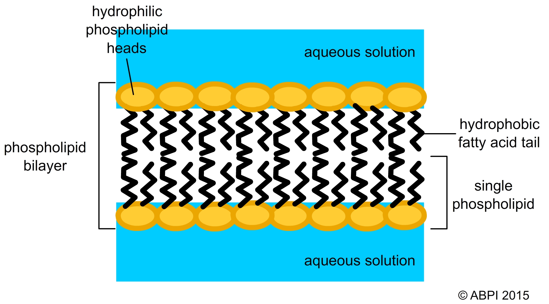
Cell membranes
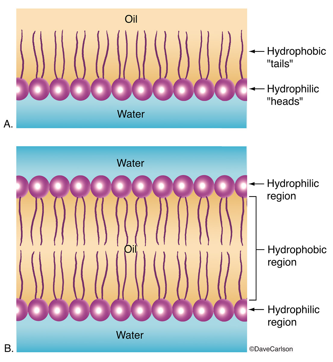
Phospholipid Layer & Bilayer Carlson Stock Art
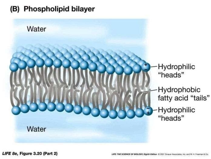
Phospholipid Bilayer Lipid Bilayer Structures & Functions
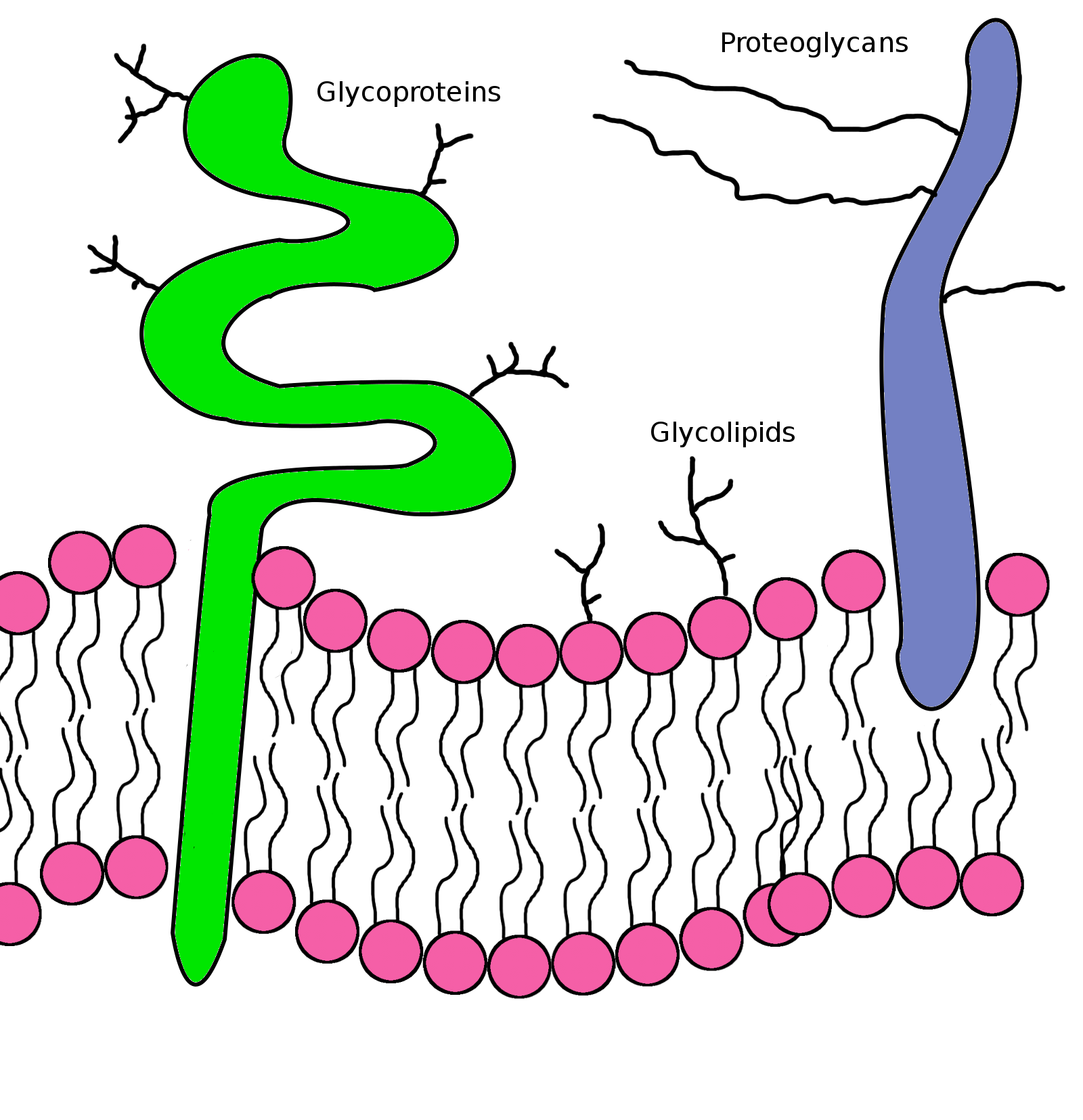
Phospholipid bilayers made easy Science is Delicious
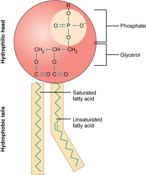
Phospholipid Bilayer Introduction, Structure and Functions
Describe the Structure of Phospholipid Bilayer ClaudiahasCarlson

How to Draw a Phospholipid Bilayer YouTube

TJ. Schematic diagram of typical membrane proteins in a biological
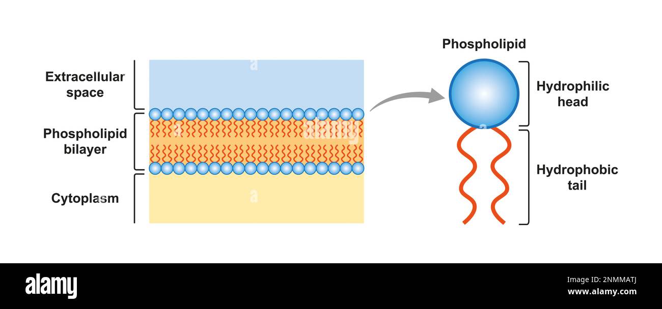
Phospholipid bilayer structure, illustration Stock Photo Alamy
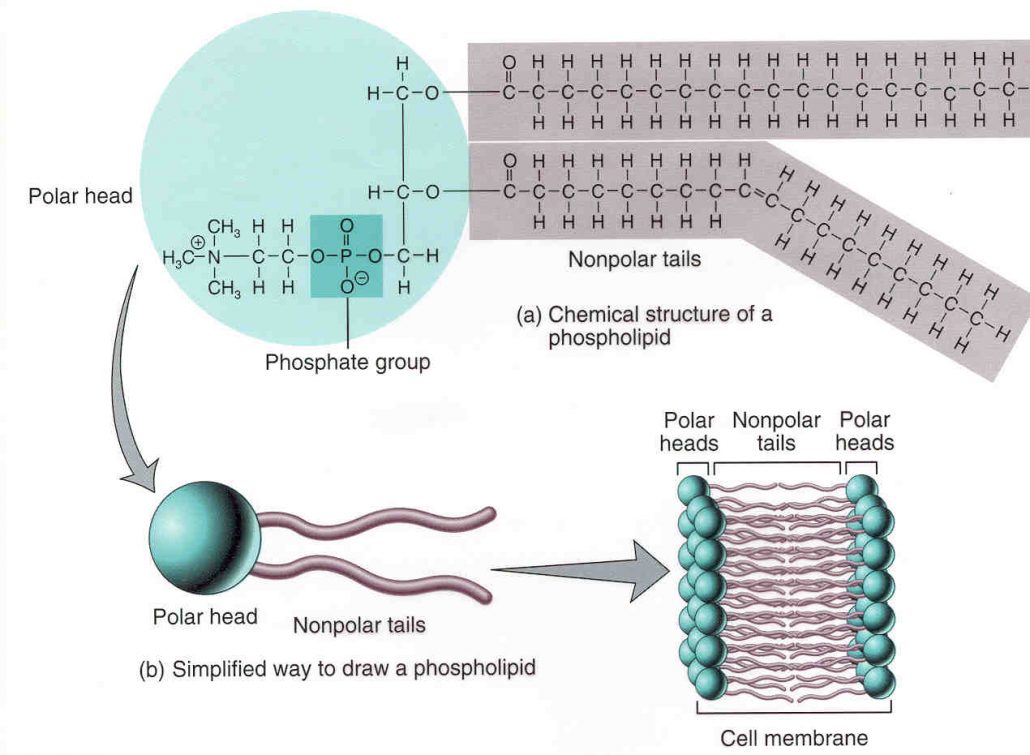
Phospholipid Bilayer Introduction, Structure and Functions
Click The Card To Flip 👆.
This Membrane Surrounds The Cell.
Web The Lipid Bilayer (Or Phospholipid Bilayer) Is A Thin Polar Membrane Made Of Two Layers Of Lipid Molecules.
Web A Phospholipid Bilayer Is Composed Of Two Layers Of Phospholipids;
Related Post:
