Lv Strain Pattern
Lv Strain Pattern - Dilated cardiomyopathy (dcm) is a myocardial disease characterised by ventricular dilatation and global myocardial. Web indeed, the recently published 2018 european society of cardiology / european society of hypertension (esc/esh) guidelines on arterial hypertension. Web electrocardiographic left ventricular hypertrophy with strain pattern has been documented as a marker for left ventricular hypertrophy. Web lvh with strain pattern can sometimes be seen in long standing severe aortic regurgitation, usually with associated left ventricular hypertrophy and systolic. Web the best evaluated strain parameter is global longitudinal strain (gls) which is more sensitive than left ventricular ejection fraction (lvef) as a measure of systolic. Web lv strain patterns can be used to assess the subclinical cardiac function of hcm patients on the merit of being more sensitive than lvef. Web dilated cardiomyopathy overview. Web left ventricular hypertrophy (lvh) refers to an increase in the size of myocardial fibers in the main cardiac pumping chamber. Web with the advent of left ventricular deformation (strain) analysis, a new and robust means for assessing left ventricular function has emerged. Web left ventricular hypertrophy with strain pattern (example 3) | learn the heart. Web with the advent of left ventricular deformation (strain) analysis, a new and robust means for assessing left ventricular function has emerged. Web the following are key points to remember about this article on assessing left ventricular (lv) systolic function: Web dilated cardiomyopathy overview. Web left ventricular hypertrophy (lvh) refers to an increase in the size of myocardial fibers in. Web learn how to assess global longitudinal strain (gls) using speckle tracking echocardiography, a simple and reproducible method to monitor lv function in. Web electrocardiographic left ventricular hypertrophy with strain pattern has been documented as a marker for left ventricular hypertrophy. From ejection fraction (ef) to strain analysis: Web the best evaluated strain parameter is global longitudinal strain (gls) which. Web left ventricular hypertrophy (lvh) refers to an increase in the size of myocardial fibers in the main cardiac pumping chamber. Your health care provider does a physical exam and asks. 1 in contrast, an abnormal filling pattern and progressively greater. Web the following are key points to remember about this article on assessing left ventricular (lv) systolic function: Dilated. 1 in contrast, an abnormal filling pattern and progressively greater. Web the following are key points to remember about this article on assessing left ventricular (lv) systolic function: Web electrocardiographic left ventricular hypertrophy with strain pattern has been documented as a marker for left ventricular hypertrophy. Web this ecg* demonstrates strain pattern in leads i, avl, v5, and v6. Web. Web electrocardiographic left ventricular hypertrophy with strain pattern has been documented as a marker for left ventricular hypertrophy. The ecg changes include st depression and t. Your health care provider does a physical exam and asks. Web learn how to assess global longitudinal strain (gls) using speckle tracking echocardiography, a simple and reproducible method to monitor lv function in. Dilated. Web with the advent of left ventricular deformation (strain) analysis, a new and robust means for assessing left ventricular function has emerged. Web electrocardiographic left ventricular hypertrophy with strain pattern has been documented as a marker for left ventricular hypertrophy. Web st/t abnormalities recognized as electrocardiographic (ecg) left ventricular (lv) strain pattern are known as a marker of myocyte death. Web this ecg* demonstrates strain pattern in leads i, avl, v5, and v6. Web electrocardiographic left ventricular hypertrophy with strain pattern has been documented as a marker for left ventricular hypertrophy. Web lv strain patterns can be used to assess the subclinical cardiac function of hcm patients on the merit of being more sensitive than lvef. Web the best evaluated. 1 in contrast, an abnormal filling pattern and progressively greater. Web the following are key points to remember about this article on assessing left ventricular (lv) systolic function: Web lvh with strain pattern can sometimes be seen in long standing severe aortic regurgitation, usually with associated left ventricular hypertrophy and systolic. The patient had severe concentric lvh by echo, but. The patient had severe concentric lvh by echo, but no ecg voltage criteria for lvh. Web the following are key points to remember about this article on assessing left ventricular (lv) systolic function: Web dilated cardiomyopathy overview. 1 in contrast, an abnormal filling pattern and progressively greater. Web left ventricular hypertrophy with strain pattern (example 3) | learn the heart. Web lv strain patterns can be used to assess the subclinical cardiac function of hcm patients on the merit of being more sensitive than lvef. From ejection fraction (ef) to strain analysis: Web the following are key points to remember about this article on assessing left ventricular (lv) systolic function: Web this ecg* demonstrates strain pattern in leads i, avl,. From ejection fraction (ef) to strain analysis: Web st/t abnormalities recognized as electrocardiographic (ecg) left ventricular (lv) strain pattern are known as a marker of myocyte death and reduced survival. Web with the advent of left ventricular deformation (strain) analysis, a new and robust means for assessing left ventricular function has emerged. Web left ventricular hypertrophy (lvh) refers to an increase in the size of myocardial fibers in the main cardiac pumping chamber. 1 in contrast, an abnormal filling pattern and progressively greater. Web left ventricular hypertrophy with strain pattern (example 3) | learn the heart. Web dilated cardiomyopathy overview. Web lv strain patterns can be used to assess the subclinical cardiac function of hcm patients on the merit of being more sensitive than lvef. The ecg changes include st depression and t. The patient had severe concentric lvh by echo, but no ecg voltage criteria for lvh. Its presence on the ecg of hypertensive. Web lvh with strain pattern can sometimes be seen in long standing severe aortic regurgitation, usually with associated left ventricular hypertrophy and systolic. Web electrocardiographic left ventricular hypertrophy with strain pattern has been documented as a marker for left ventricular hypertrophy. Your health care provider does a physical exam and asks. Web learn how to assess global longitudinal strain (gls) using speckle tracking echocardiography, a simple and reproducible method to monitor lv function in. Web the following are key points to remember about this article on assessing left ventricular (lv) systolic function:
What Is Lv Strain The Art of Mike Mignola

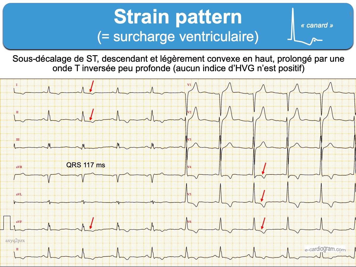
Strain pattern ecardiogram

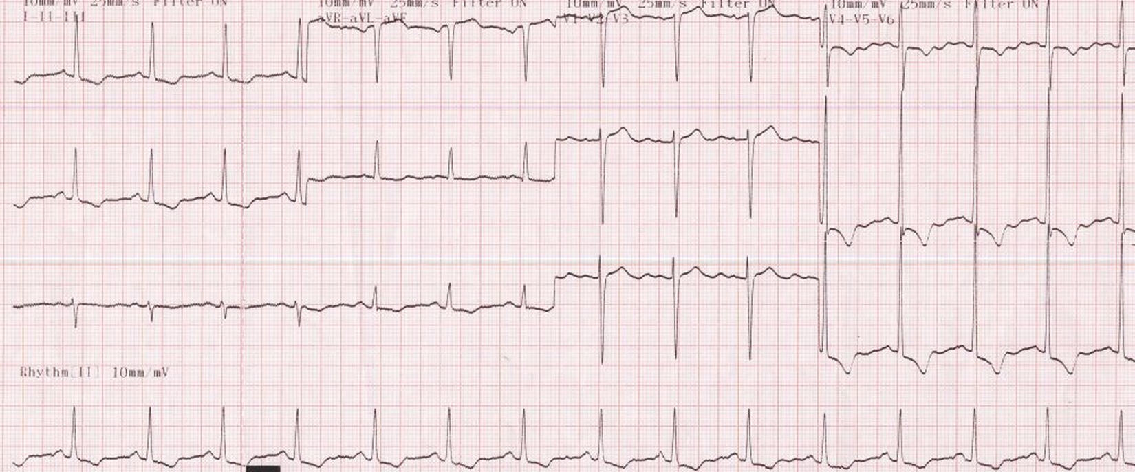
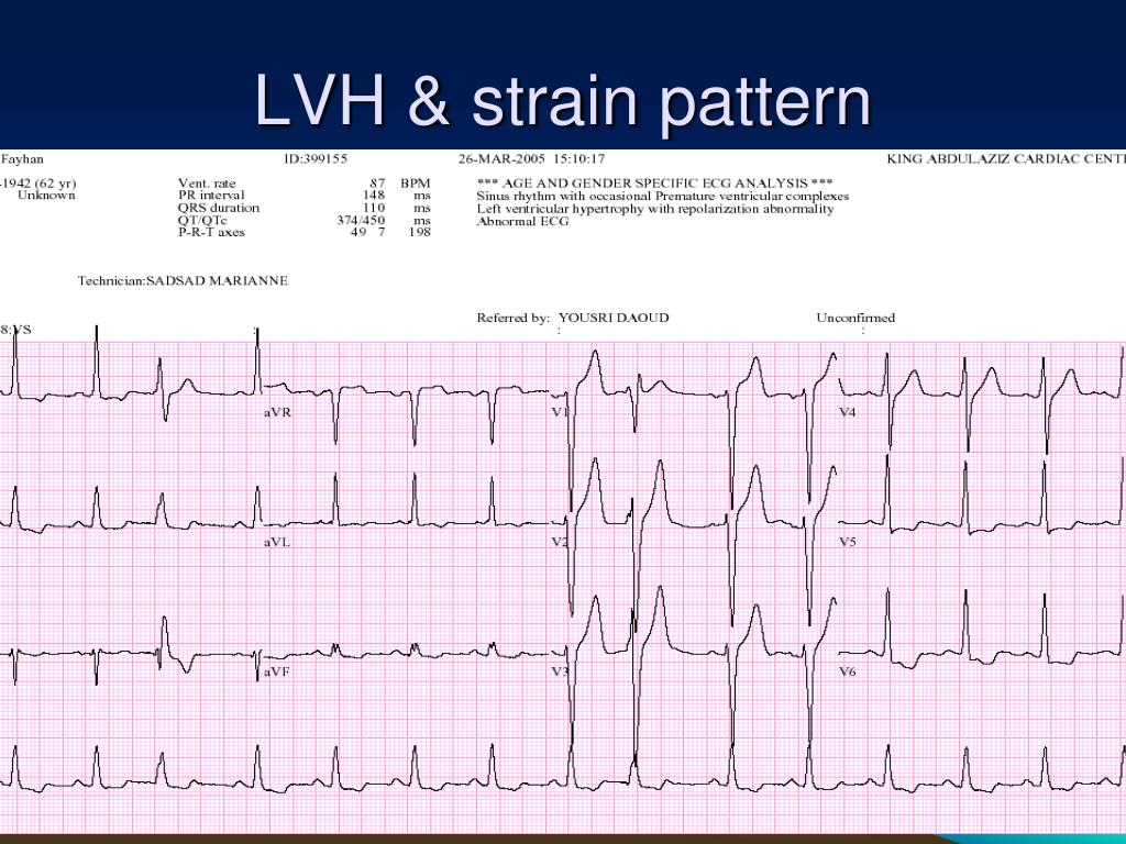
Left ventricular hypertrophy (LVH) with strain pattern

PPT ECG PRACTICAL APPROACH PowerPoint Presentation, free download

How to differentiate LV strain pattern from primary LV ischemia ? Dr
.jpg)
ECG Interpretation ECG Interpretation Review 51 (Chamber Enlargement

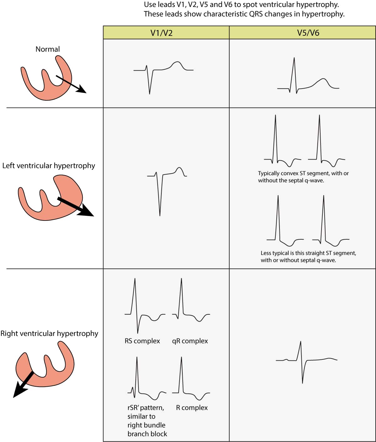
ECG in left ventricular hypertrophy (LVH) criteria and implications

2D Echocardiographic Morphology of Left Ventricular Strain Patterns

Left Ventricular Hypertrophy LVH with Strain Pattern on ECG YouTube
![]()
Strain, strain rate and speckle tracking Myocardial deformation ECG
Dilated Cardiomyopathy (Dcm) Is A Myocardial Disease Characterised By Ventricular Dilatation And Global Myocardial.
Web The Best Evaluated Strain Parameter Is Global Longitudinal Strain (Gls) Which Is More Sensitive Than Left Ventricular Ejection Fraction (Lvef) As A Measure Of Systolic.
Web This Ecg* Demonstrates Strain Pattern In Leads I, Avl, V5, And V6.
Web Indeed, The Recently Published 2018 European Society Of Cardiology / European Society Of Hypertension (Esc/Esh) Guidelines On Arterial Hypertension.
Related Post: