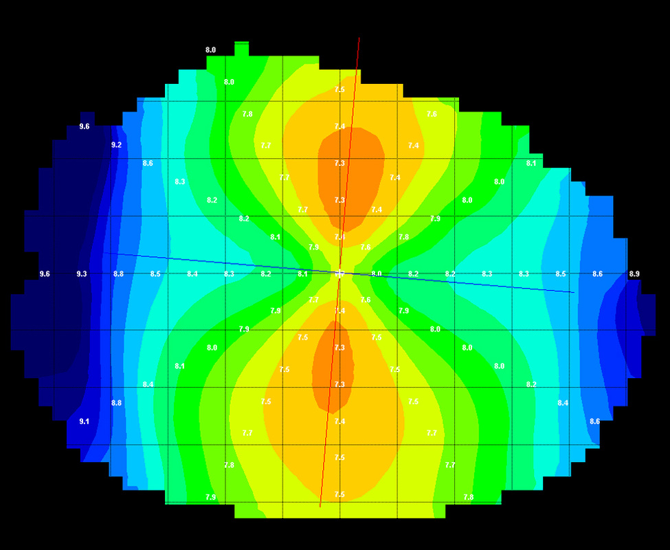
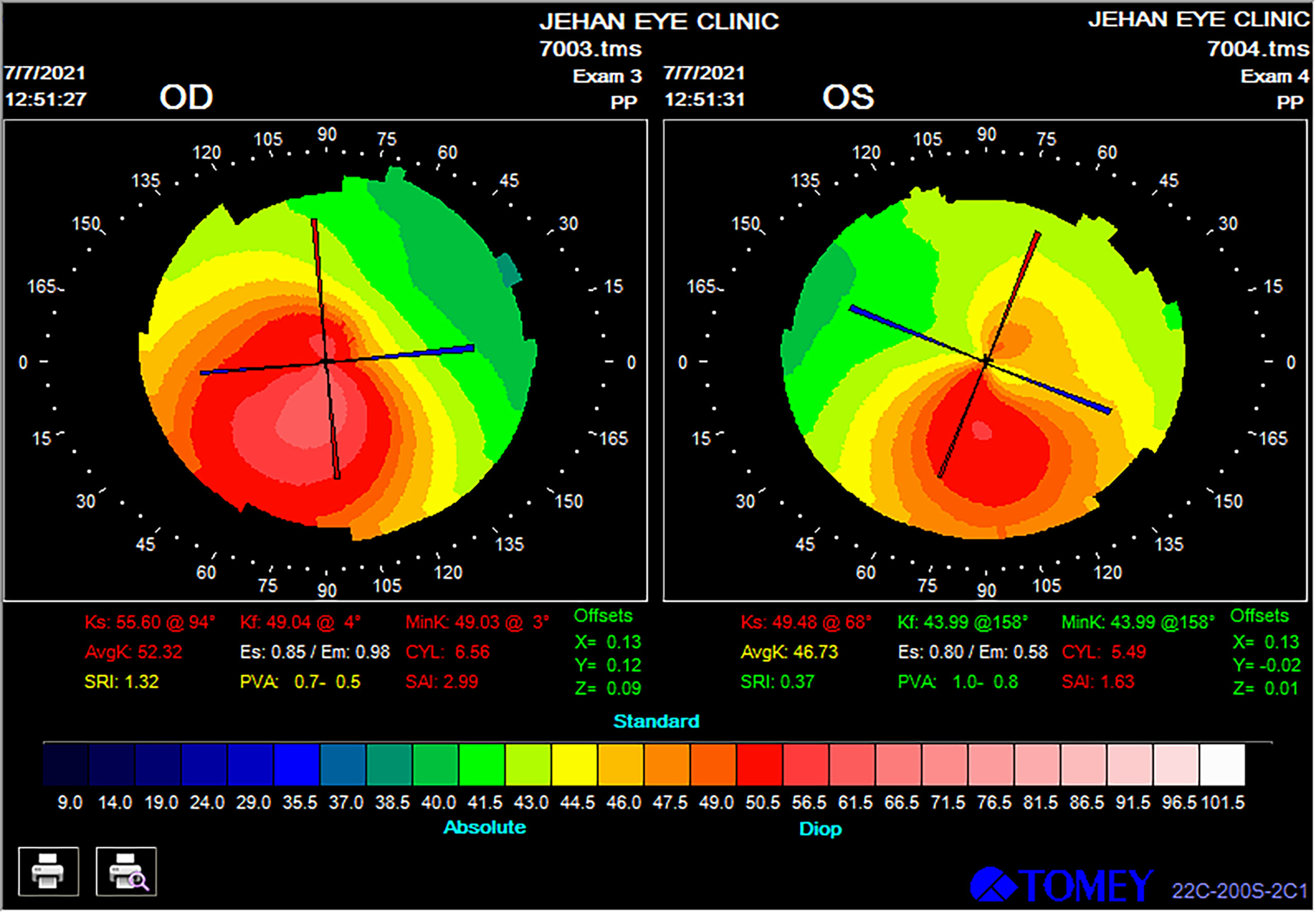
Keratoconus Topography Patterns
Keratoconus Topography Patterns - Web keratoconus is a common progressive corneal disorder that can be associated with significant ocular morbidity. Web published april 15, 2024. Diagnosis of keratoconus has greatly improved from simple clinical diagnosis with the advent of better diagnostic devices like corneal. Web oct corneal and epithelial thickness map parameters and patterns can be used in conjunction with topography to improve keratoconus screening. Web topographic pattern recognition: Web topography is evaluated using placido disk patterns or mires reflected off of the tear film of the anterior cornea and converted to color scales. Web topography of a patient with a pattern diagnosis revealing a classic asymmetric bowtie pattern with skewed radial axis consistent with keratoconus. Fitting rigid gas permeable contact lenses (rgp cls) in keratoconic patients is the most common visual rehabilitation option to improve. Signs of early disease can be subtle and easy to. Corneal topography is a noninvasive eye exam that analyzes both qualitatively and quantitatively the morphology of the cornea [ 1 ], providing its. This asymmetry manifests in irregular curvature patterns, focal asymmetric corneal thinning, focal. Web 8) keratoconus prediction index (kpi) describes the percentage probability of keratoconus by analyzing the topographic data of anterior corneal surface. Web christopher kent, senior editor. Corneal topography is a noninvasive eye exam that analyzes both qualitatively and quantitatively the morphology of the cornea [ 1 ], providing. Diagnosis of keratoconus has greatly improved from simple clinical diagnosis with the advent of better diagnostic devices like corneal. Web topographic pattern recognition: Web published april 15, 2024. Web the hallmark of corneal ectatic disorders is asymmetry: Early signs of keratoconus include: Web in this study, the shape of corneal distortion manifested by the keratoconus has been detected by extracting a pattern of corneal irregularities from the given. Web oct corneal and epithelial thickness map parameters and patterns can be used in conjunction with topography to improve keratoconus screening. Keratoconus is defined as a chronic, noninflammatory ectatic disorder of the cornea, characterized. Web a combination of multiple imaging tools, including corneal topography, tomography, scheimpflug imaging, anterior segment optical coherence tomography, and. In incipient cases, however, the use of a single parameter to diagnose keratoconus is. Web topography is evaluated using placido disk patterns or mires reflected off of the tear film of the anterior cornea and converted to color scales. Three different. Web christopher kent, senior editor. The roles of topography and tomography. Because the image is generated off. Corneal topography is a noninvasive eye exam that analyzes both qualitatively and quantitatively the morphology of the cornea [ 1 ], providing its. Keratoconus is defined as a chronic, noninflammatory ectatic disorder of the cornea, characterized by steepening, apical. Web a combination of multiple imaging tools, including corneal topography, tomography, scheimpflug imaging, anterior segment optical coherence tomography, and. Web the hallmark of corneal ectatic disorders is asymmetry: Web christopher kent, senior editor. Web topographic pattern recognition: Signs of early disease can be subtle and easy to. Web keratoconus is a common progressive corneal disorder that can be associated with significant ocular morbidity. Web topography of a patient with a pattern diagnosis revealing a classic asymmetric bowtie pattern with skewed radial axis consistent with keratoconus. Keratoconus is defined as a chronic, noninflammatory ectatic disorder of the cornea, characterized by steepening, apical. Signs of early disease can be. In incipient cases, however, the use of a single parameter to diagnose keratoconus is. Web the hallmark of corneal ectatic disorders is asymmetry: Web keratoconus is a common progressive corneal disorder that can be associated with significant ocular morbidity. Web corneal topography is the primary diagnostic tool for keratoconus detection. Keratoconus is defined as a chronic, noninflammatory ectatic disorder of. Web in this study, the shape of corneal distortion manifested by the keratoconus has been detected by extracting a pattern of corneal irregularities from the given. Fitting rigid gas permeable contact lenses (rgp cls) in keratoconic patients is the most common visual rehabilitation option to improve. The roles of topography and tomography. Diagnosis of keratoconus has greatly improved from simple. In incipient cases, however, the use of a single parameter to diagnose keratoconus is. Web keratometry/computerized topography/computerized tomography/ultrasound pachymetry; Three different imaging modalities serve their own purposes for. Because the image is generated off. Various corneal imaging techniques have. Web a combination of multiple imaging tools, including corneal topography, tomography, scheimpflug imaging, anterior segment optical coherence tomography, and. In incipient cases, however, the use of a single parameter to diagnose keratoconus is. Because the image is generated off. Web keratometry/computerized topography/computerized tomography/ultrasound pachymetry; Early signs of keratoconus include: Various corneal imaging techniques have. Web the hallmark of corneal ectatic disorders is asymmetry: Keratoconus is defined as a chronic, noninflammatory ectatic disorder of the cornea, characterized by steepening, apical. Signs of early disease can be subtle and easy to. Web corneal topography is the primary diagnostic tool for keratoconus detection. Web corneal topography is an important tool for detecting corneal morphology, enabling both correct classification of keratoconus (kc) and detection of suspicious or. Corneal topography and tomography provide valuable information about the corneal curvature. Web topography of a patient with a pattern diagnosis revealing a classic asymmetric bowtie pattern with skewed radial axis consistent with keratoconus. Web oct corneal and epithelial thickness map parameters and patterns can be used in conjunction with topography to improve keratoconus screening. Fitting rigid gas permeable contact lenses (rgp cls) in keratoconic patients is the most common visual rehabilitation option to improve. Web topography is evaluated using placido disk patterns or mires reflected off of the tear film of the anterior cornea and converted to color scales.
Proposed classification for topographic patterns seen after

Sagittal curvature map of a keratoconus cornea obtained by a rotating

Topography Keratoconus Contact Lens Fitting

Keratoconus diagnosis, treatment and management in Vancouver, British

Keratoconus Corneal Topography

Corneal Topography EyeWiki

Axial curvature maps of corneal topography pattern of mild to moderate

Corneal topography in a keratoconus eye. Download Scientific Diagram

Topography Keratoconus Contact Lens Fitting

Imaging modalities in keratoconus Matalia H, Swarup R Indian J Ophthalmol
Three Different Imaging Modalities Serve Their Own Purposes For.
The Roles Of Topography And Tomography.
Corneal Topography Is A Noninvasive Eye Exam That Analyzes Both Qualitatively And Quantitatively The Morphology Of The Cornea [ 1 ], Providing Its.
Web Keratoconus Is A Common Progressive Corneal Disorder That Can Be Associated With Significant Ocular Morbidity.
Related Post: