Homogeneous Speckled Ana Pattern
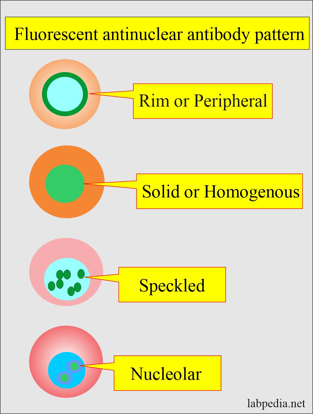
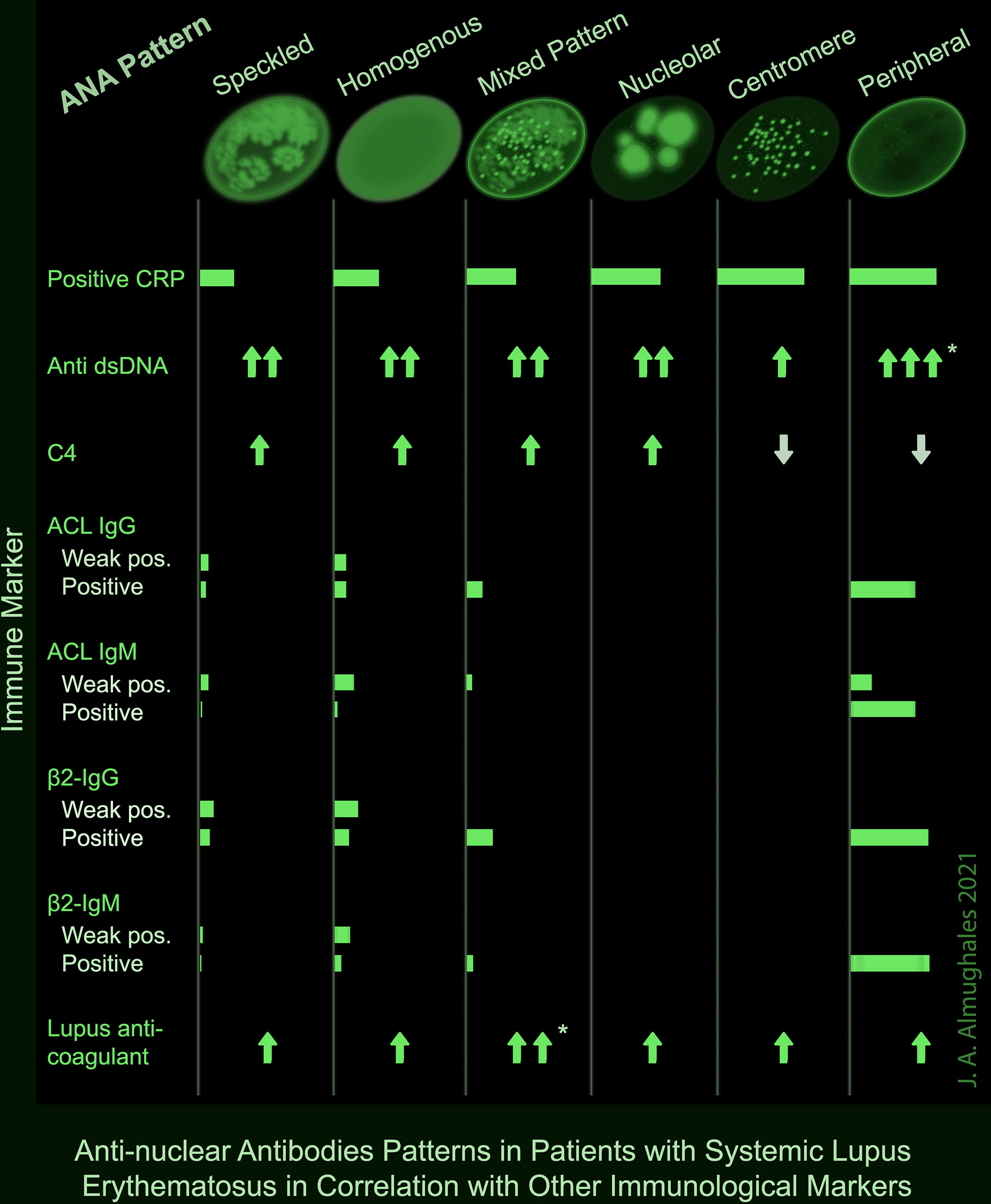
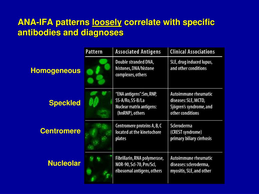
Homogeneous Speckled Ana Pattern - Your immune system normally makes antibodies to help you fight infection. Some, but not all labs will report a titre above 1:160 as positive. Web this retrospective study showed that speckled followed by homogeneous ana patterns were predominant accounting for 52.1 and 35.2% of the patients. The commonly recognized patterns include: Web an ana test was performed, with a strongly positive (30 iu/ml or >1:1 280) homogeneous pattern. Ana pattern is most commonly speckled, followed by centromeric. Antibodies that attack healthy proteins within the cell nucleus are called antinuclear antibodies (anas). An ana test detects antinuclear antibodies (ana) in your blood. Antibodies are proteins that are made as part of an immune response. Fine and coarse speckles of ana staining Common ana pattern is speckled; The pattern refers to the distribution of staining produced by autoantibodies reacting with antigens in the cells. Patterns that are reported include, homogeneous, speckled, centromere, and others. Web the presence of ana with a homogeneous & speckled (hs) pattern was significantly associated with the absence of cancer ( < 0.01). Web ana titers and patterns. Dipstick urinalysis showed moderate blood and ++ protein. A homogenous staining pattern means the entire nucleus is stained with ana. The entire nucleus is stained with ana. Web the patterns seen are as follows: Web a health care provider may request that a patient have a test for antinuclear antibodies (ana) as part of an evaluation for possible autoimmune disease. A speckled staining pattern means fine, coarse speckles of ana are present. When antibodies to dna and deoxyribonucleoprotein are present (rim and homogenous pattern), there may be interference with the detection of speckled pattern. This is the most common pattern and can be seen with any autoimmune disease. In sjögren syndrome there will often be a speckled pattern; It’s the. For this test, we use a. Web is the ana pattern suggestive of a specific disease? Normally, the immune system responds to an infection by producing large numbers of antibodies to fight bacteria or. In sjögren syndrome there will often be a speckled pattern; It is found in many disease states, including sle and scleroderma. Web antinuclear antibody panel (ana test) uses. Homogenous staining can result from antibodies to dna and histones. Web is the ana pattern suggestive of a specific disease? Homogeneous and regular fluorescence across all nucleoplasm. The commonly recognized patterns include: Titres are reported in ratios, most often 1:40, 1:80, 1:160, 1:320, and 1:640. Antibodies are proteins that are made as part of an immune response. Homogeneous and regular fluorescence across all nucleoplasm. A homogenous staining pattern means the entire nucleus is stained with ana. It is found in many disease states, including sle and scleroderma. The pattern refers to the distribution of staining produced by autoantibodies reacting with antigens in the cells. Homogenous staining can result from antibodies to dna and histones. The commonly recognized patterns include: Homogeneous — staining is even in the entire nucleus and is commonly found in people with sle and discoid lupus erythematosus (dle). The level or titer and the. Mitotic cells (metaphase, anaphase, and telophase) have the chromatin mass intensely stained in a homogeneous hyaline fashion. Some, but not all labs will report a titre above 1:160 as positive. This is the most common pattern and can be seen with any autoimmune disease. Common ana pattern is speckled; The ana pattern showed several associations with other immune markers that. Web the presence of ana with a homogeneous & speckled (hs) pattern was significantly associated with the absence of cancer ( < 0.01). The commonly recognized patterns include: Web antinuclear antibody panel (ana test) uses. Web a health care provider may request that a patient have a test for antinuclear antibodies (ana) as part of an evaluation for possible autoimmune. Total nuclear fluorescence due to an antibody directed against dna or histone proteins. Web is the ana pattern suggestive of a specific disease? Web this retrospective study showed that speckled followed by homogeneous ana patterns were predominant accounting for 52.1 and 35.2% of the patients. Ana pattern is most commonly speckled, followed by centromeric. Mitotic cells (metaphase, anaphase, and telophase). At times, laboratories testing ana also report a “pattern”. Homogeneous and regular fluorescence across all nucleoplasm. Web is the ana pattern suggestive of a specific disease? A homogenous (diffuse) pattern appears as total nuclear fluorescence and is common in people with systemic lupus. Antibodies that attack healthy proteins within the cell nucleus are called antinuclear antibodies (anas). Titres are reported in ratios, most often 1:40, 1:80, 1:160, 1:320, and 1:640. Ana pattern is most commonly speckled, followed by centromeric. The ana pattern showed several associations with other immune markers that are documented to have significant clinical implications in sle. The nucleoli maybe stained or not stained depending on cell substrate. Web the patterns seen are as follows: This is the most common pattern and can be seen with any autoimmune disease. Web an ana test was performed, with a strongly positive (30 iu/ml or >1:1 280) homogeneous pattern. Total nuclear fluorescence due to an antibody directed against dna or histone proteins. The level or titer and the pattern. A homogeneous/peripheral pattern reflects antibodies to histone/dsdna/chromatin, whereas many other specificities found in systemic rheumatic diseases show speckled patterns of. The commonly recognized patterns include:
Antinuclear Factor (ANF), Antinuclear Antibody (ANA) and Its
.jpg)
ANA Mixed pattern University of Birmingham

Frontiers AntiNuclear Antibodies Patterns in Patients With Systemic

Biochemistry, Antinuclear Antibodies (ANA) StatPearls NCBI Bookshelf

Ana Titer 1 160 Speckled Pattern Chumado

ANA Patterns

DFS70 antibodies biomarkers for the exclusion of ANAassociated

Common ANA patterns by IIF a, negative sample; b, homogeneous; c

ANA Patterns

Ana With Speckled Pattern Chumado
An Ana Test Detects Antinuclear Antibodies (Ana) In Your Blood.
It’s The Most Common Type Of Staining Pattern.
The Pattern Refers To The Distribution Of Staining Produced By Autoantibodies Reacting With Antigens In The Cells.
Your Immune System Normally Makes Antibodies To Help You Fight Infection.
Related Post: