Hep2 Speckled Pattern
Hep2 Speckled Pattern - Certain diseases are more likely to have certain. Titres are reported in ratios, most often 1:40, 1:80, 1:160, 1:320, and 1:640. Homogenous, speckled, centromere, nucleolar, and nuclear dots. Web the gold standard method for ana determination is indirect immunofluorescence (iif) on the human laryngeal epidermoid carcinoma cell line type 2. Assess antinuclear antibody positivity, titers, and patterns display differences in various sard,. (a) icap classification chart in use from 2015 to 2021. Web a positive ana test is usually reported as both a ratio (called a titer) and a pattern, such as smooth or speckled. Icap was initiated as a workshop aiming. These patterns are the result of autoantibody binding to specific. When active, usually a homogenous pattern on ana or less commonly speckled, rim, or. Homogenous, speckled, centromere, nucleolar, and nuclear dots. Web in august 2014, the international consensus on antinuclear antibody pattern meeting made clear that cytoplasmic patterns were categorized as a major group and. Titres are reported in ratios, most often 1:40, 1:80, 1:160, 1:320, and 1:640. Certain diseases are more likely to have certain. (a) icap classification chart in use from 2015. Certain diseases are more likely to have certain. A survey of laboratory performance, pattern recognition and interpretation. Web in august 2014, the international consensus on antinuclear antibody pattern meeting made clear that cytoplasmic patterns were categorized as a major group and. Web the gold standard method for ana determination is indirect immunofluorescence (iif) on the human laryngeal epidermoid carcinoma cell. Ana test results are most often reported in 2 parts: Assess antinuclear antibody positivity, titers, and patterns display differences in various sard,. Web the gold standard method for ana determination is indirect immunofluorescence (iif) on the human laryngeal epidermoid carcinoma cell line type 2. (a) icap classification chart in use from 2015 to 2021. Web the major pattern in hc. Web the gold standard method for ana determination is indirect immunofluorescence (iif) on the human laryngeal epidermoid carcinoma cell line type 2. Web a positive ana test is usually reported as both a ratio (called a titer) and a pattern, such as smooth or speckled. Titres are reported in ratios, most often 1:40, 1:80, 1:160, 1:320, and 1:640. (a) icap. Icap was initiated as a workshop aiming. Ana test results are most often reported in 2 parts: Assess antinuclear antibody positivity, titers, and patterns display differences in various sard,. Homogenous, speckled, centromere, nucleolar, and nuclear dots. Web a positive ana test is usually reported as both a ratio (called a titer) and a pattern, such as smooth or speckled. Web the gold standard method for ana determination is indirect immunofluorescence (iif) on the human laryngeal epidermoid carcinoma cell line type 2. Web the most frequent ana pattern in both groups was the nuclear fine speckled pattern, which occurred at lower titer in healthy individuals than in patients with ards (p<0.001). Web a positive ana test is usually reported as. Web in august 2014, the international consensus on antinuclear antibody pattern meeting made clear that cytoplasmic patterns were categorized as a major group and. Icap was initiated as a workshop aiming. Assess antinuclear antibody positivity, titers, and patterns display differences in various sard,. Web the gold standard method for ana determination is indirect immunofluorescence (iif) on the human laryngeal epidermoid. Web the major pattern in hc was ac‐2 (12.2%). Titres are reported in ratios, most often 1:40, 1:80, 1:160, 1:320, and 1:640. Web a positive ana test is usually reported as both a ratio (called a titer) and a pattern, such as smooth or speckled. Assess antinuclear antibody positivity, titers, and patterns display differences in various sard,. The level or. The level or titer and the pattern. A survey of laboratory performance, pattern recognition and interpretation. Web in august 2014, the international consensus on antinuclear antibody pattern meeting made clear that cytoplasmic patterns were categorized as a major group and. These patterns are the result of autoantibody binding to specific. Web a homogeneous/peripheral pattern reflects antibodies to histone/dsdna/chromatin, whereas many. (a) icap classification chart in use from 2015 to 2021. Ana test results are most often reported in 2 parts: The level or titer and the pattern. Web a homogeneous/peripheral pattern reflects antibodies to histone/dsdna/chromatin, whereas many other specificities found in systemic rheumatic diseases show speckled. Assess antinuclear antibody positivity, titers, and patterns display differences in various sard,. When active, usually a homogenous pattern on ana or less commonly speckled, rim, or. These patterns are the result of autoantibody binding to specific. Ana test results are most often reported in 2 parts: Web the most frequent ana pattern in both groups was the nuclear fine speckled pattern, which occurred at lower titer in healthy individuals than in patients with ards (p<0.001). A survey of laboratory performance, pattern recognition and interpretation. Titres are reported in ratios, most often 1:40, 1:80, 1:160, 1:320, and 1:640. Icap was initiated as a workshop aiming. (a) icap classification chart in use from 2015 to 2021. Web welcome to anapatterns.org, the official website for the international consensus on antinuclear antibody (ana) patterns (icap). The level or titer and the pattern. Web a homogeneous/peripheral pattern reflects antibodies to histone/dsdna/chromatin, whereas many other specificities found in systemic rheumatic diseases show speckled. Certain diseases are more likely to have certain. Web a positive ana test is usually reported as both a ratio (called a titer) and a pattern, such as smooth or speckled. Web the gold standard method for ana determination is indirect immunofluorescence (iif) on the human laryngeal epidermoid carcinoma cell line type 2.
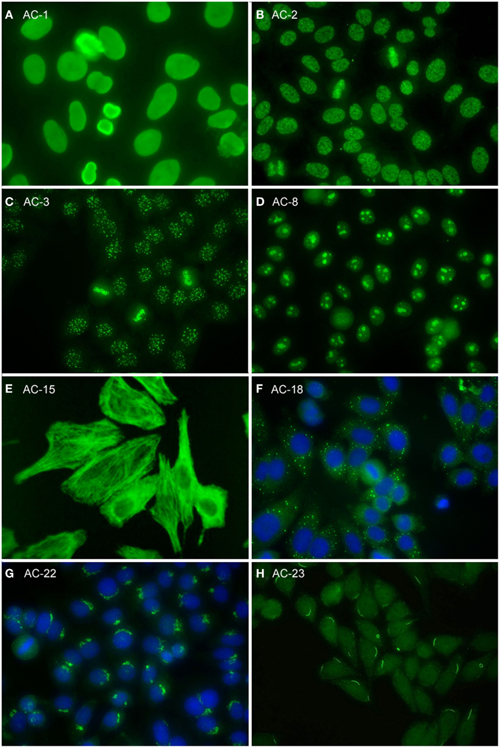
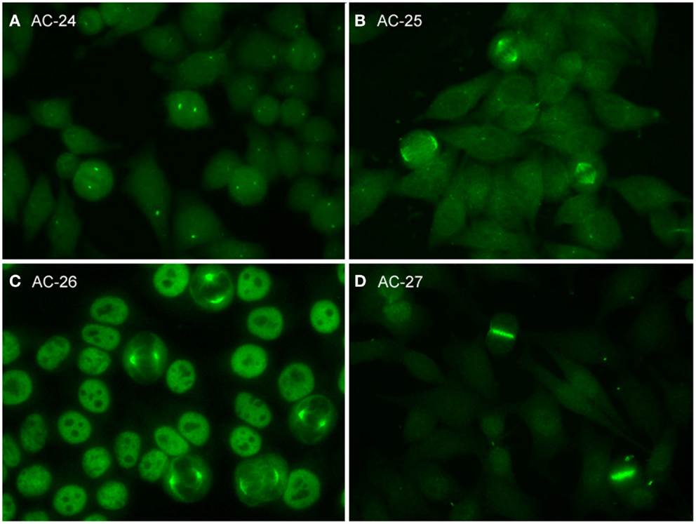
Frontiers Report of the First International Consensus on Standardized

Figure 1 from The Clinical Significance of the Dense Fine Speckled

Representative images of selected major HEp2 cell patterns. (A

HEp2 staining patterns 1) Homogeneous 2) Nucleolar 3) Coarse Speckled

Figure 3 from The Clinical Significance of the Dense Fine Speckled

IIF on Hep2 cells speckled pattern of varying intensity (antiPCNA

IIF on Hep2 cells speckled pattern (antiRNP antibodies). Dilution 1

International consensus on antinuclear antibody patterns definition of

Frontiers Report of the First International Consensus on Standardized

Finer speckled nuclear pattern and occasional HEp2 cells with PCNA
Web The Major Pattern In Hc Was Ac‐2 (12.2%).
Homogenous, Speckled, Centromere, Nucleolar, And Nuclear Dots.
Assess Antinuclear Antibody Positivity, Titers, And Patterns Display Differences In Various Sard,.
Web In August 2014, The International Consensus On Antinuclear Antibody Pattern Meeting Made Clear That Cytoplasmic Patterns Were Categorized As A Major Group And.
Related Post: