Heart Anatomy Draw
Heart Anatomy Draw - The muscular wall of the heart has three layers. The medical information on this site is provided as an information resource only, and is not to be used or relied on for any. This exercise will help you to identify your weak spots, so you’ll know which heart structures you need to spend more time. Perfect the pencil drawing before moving on to the pen. To find a good diagram, go to google images, and type in the internal structure of the human heart. This interactive atlas of human heart anatomy is based on medical illustrations and cadaver photography. Right, left, superior, and inferior: Next, draw the right atrium and the superior vena cava. Web to draw an anatomical heart requires a lot of attention and patience. Pencil sketch of the human heart embark on a journey into the beauty of human anatomy with a mesmerizing pencil sketch of the human heart. This website is aimed to be an easily accessible web based app that features an on demand high fidelity rendering of the human heart to use while on rounds, in teaching conferences, or by bedside. This is the superior vena cava. The position of the heart in the torso between the vertebrae and sternum (see figure 19.2 for the position. Xxxl very detailed human heart. The position of the heart in the torso between the vertebrae and sternum (see figure 19.2 for the position of the heart within the thorax) allows for individuals to apply an emergency technique known as cardiopulmonary resuscitation (cpr) if the heart of a patient should stop. The intricate anatomy of the heart can be challenging. The muscular wall of the heart has three layers. To find a good diagram, go to google images, and type in the internal structure of the human heart. The heart wall is composed of three layers. Web the surfaces and borders of the heart. Web heart drawing realistic anatomical beauty: For the atrium, enclose an irregular form at the junction of the ventricle and the aortic arch. Xxxl very detailed human heart. By applying pressure with the flat portion of one hand on the. The epicardium covers the heart, wraps around the roots of the great blood vessels, and adheres the heart wall to a protective sac. Your brain and. The middle layer is the myocardium. The tutorial can be done in sections, so perhaps take a break if you need to. Right, left, superior, and inferior: Web worksheet showing unlabelled heart diagrams. This artwork delves into the depths of medical illustration, celebrating the complexity and elegance of the heart's form. Location of heart in the thorax. The medical information on this site is provided as an information resource only, and is not to be used or relied on for any. Web in this lecture, dr mike shows the two best ways to draw and label the heart! Web the heart has three layers. The heart wall is composed of three. Your heart contains four muscular sections ( chambers) that briefly hold blood before moving it. Web drawing the heart anatomy can be challenging, but with practice and dedication, it can become easier over time! By applying pressure with the flat portion of one hand on the. Web drawing internal anatomy of the heart. This thick layer is the muscle that. The valves of the heart. This artwork delves into the depths of medical illustration, celebrating the complexity and elegance of the heart's form. Web heart, organ that serves as a pump to circulate the blood. The heart is located within the thoracic cavity, medially between the lungs in the mediastinum. This is the superior vena cava. Create a curved shape similar to an acorn or apple’s bottom half. This artwork delves into the depths of medical illustration, celebrating the complexity and elegance of the heart's form. The right margin is the small section of the right atrium that extends between the superior and inferior vena cava. Web still, the offscreen heart attack death of george sr.. Web heart drawing realistic anatomical beauty: For the atrium, enclose an irregular form at the junction of the ventricle and the aortic arch. Web the heart has three layers. The middle layer is the myocardium. From the openstax anatomy and physiology book. The medical information on this site is provided as an information resource only, and is not to be used or relied on for any. Electrical impulses make your heart beat, moving blood through these chambers. Web the surfaces and borders of the heart. The muscular wall of the heart has three layers. Above this, draw a narrow vertical oval along the arch, and connect this to the atrium using a curved line. The user can show or hide the anatomical labels which provide a useful tool to create illustrations perfectly adapted for teaching. Emojis help to capture and illustrate our mood without even having to express ourselves verbally. Web anatomy of the heart made easy along with the blood flow through the cardiac structures, valves, atria, and ventricles. Use a pen or pencil to draw the heart's main body. The heart wall is composed of three layers. The middle layer is the myocardium. Find a piece of paper and something to draw with. A drawing of the anatomy of the opened normal heart, with english labels. It may be a straight tube, as in spiders and annelid worms, or a somewhat more elaborate structure with one or more receiving chambers (atria) and a main pumping chamber (ventricle), as in mollusks. Web muscle and tissue make up this powerhouse organ. The heart is located within the thoracic cavity, medially between the lungs in the mediastinum.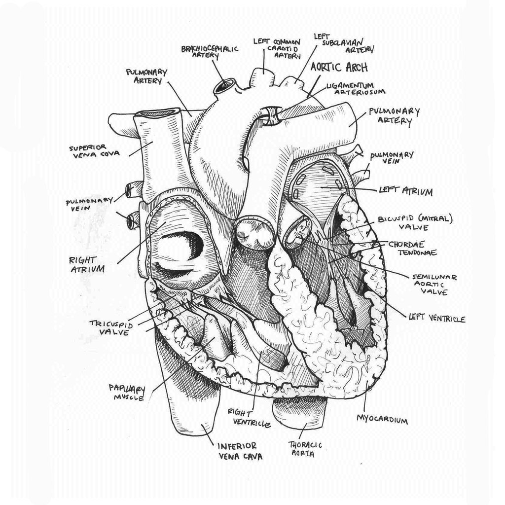
Anatomical Drawing Heart at GetDrawings Free download

How to Draw the Internal Structure of the Heart (with Pictures)

How to Draw the Internal Structure of the Heart 13 Steps
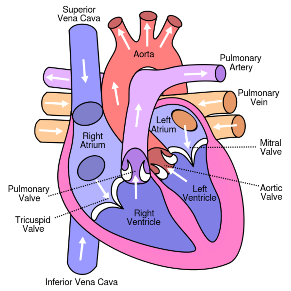
Learn About the Heart and Circulatory System for Kids HubPages
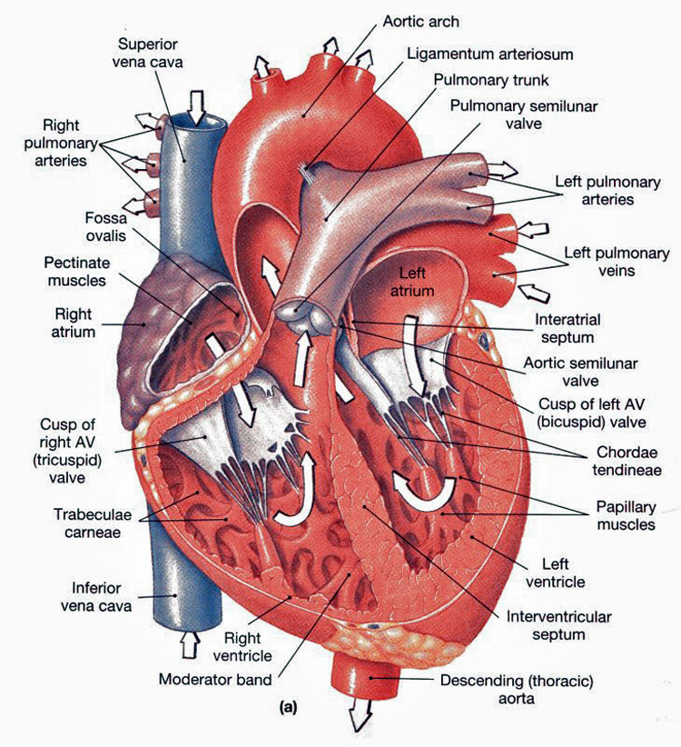
Heart Anatomy chambers, valves and vessels Anatomy & Physiology
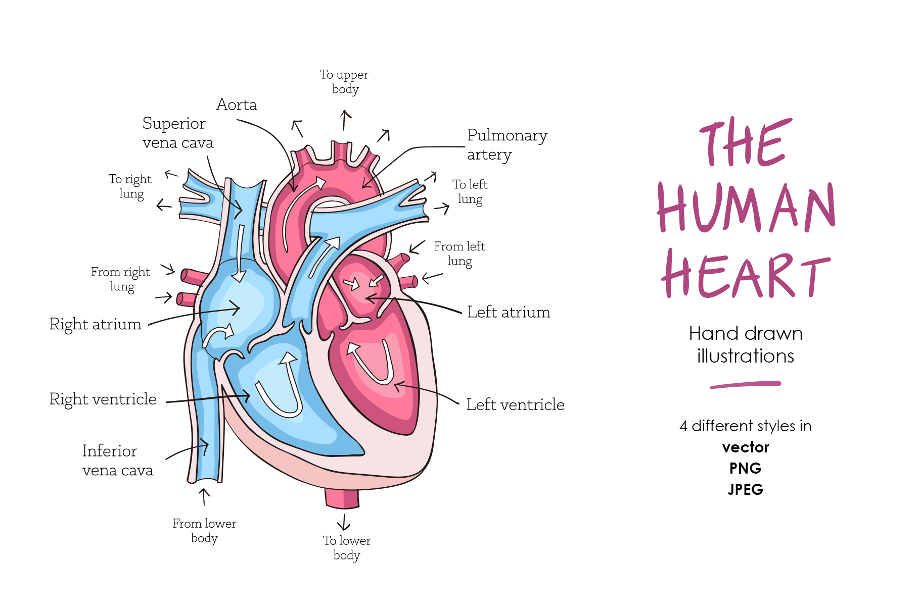
Human heart anatomy (274491) Illustrations Design Bundles
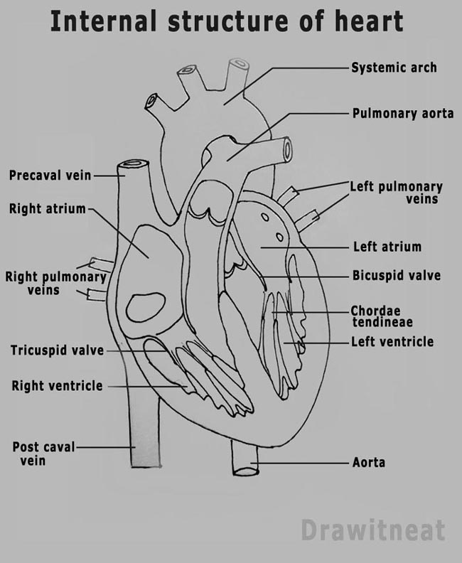
DRAW IT NEAT How to draw human heart labeled
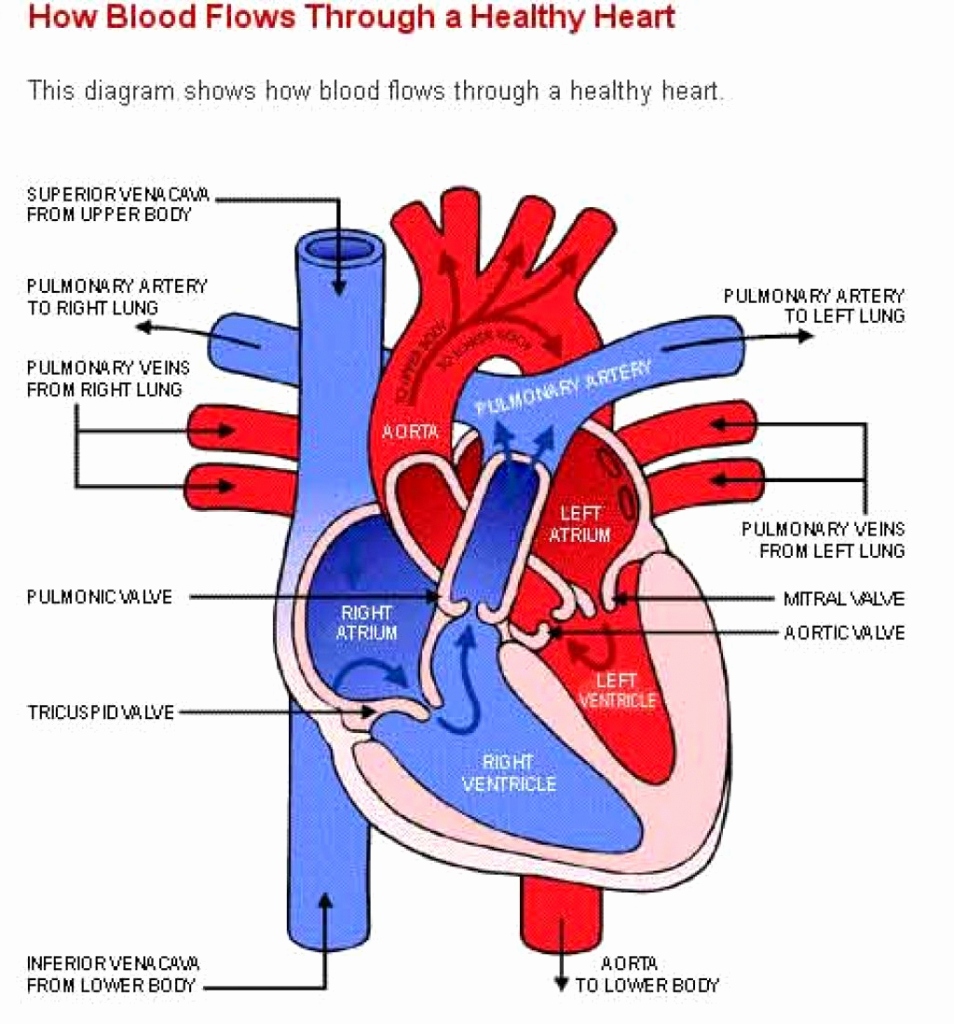
Human Heart Drawing Simple at Explore collection
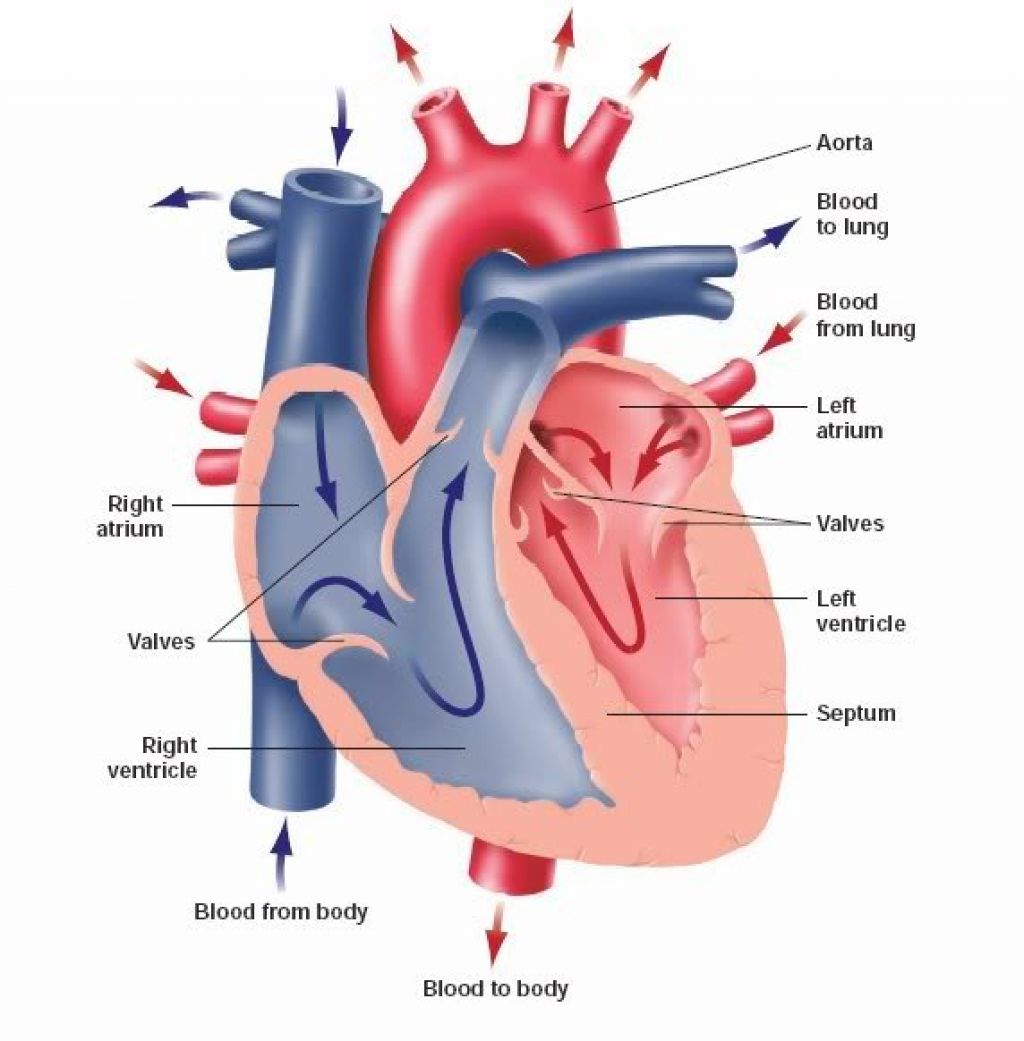
When one teaches, two learn. The heart and the circulatory system
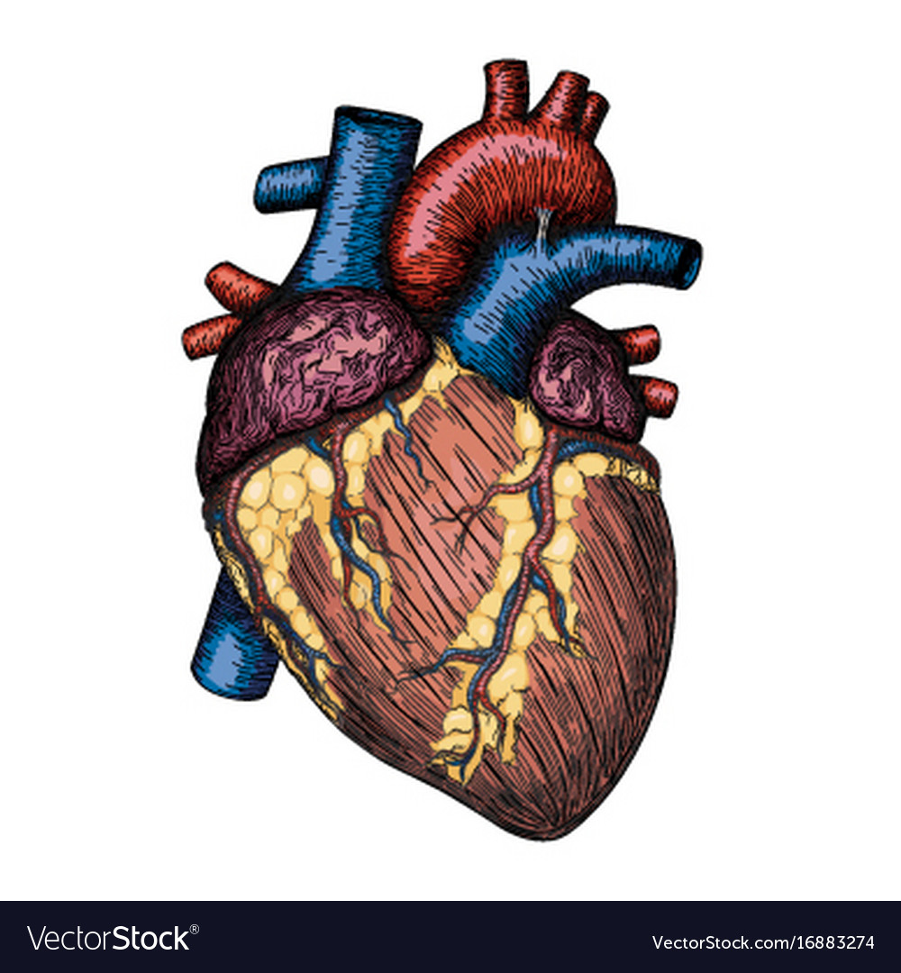
Human heart hand drawn anatomical sketch Vector Image
The Human Heart Is A Complex Organ With Many Parts And Intricate Details, Which Can Make It Difficult To Draw Accurately.
The Outermost Layer Is The Epicardium (Or Visceral Pericardium).
Web The Heart Has Three Layers.
The Right Margin Is The Small Section Of The Right Atrium That Extends Between The Superior And Inferior Vena Cava.
Related Post: