Fana Staining Patterns
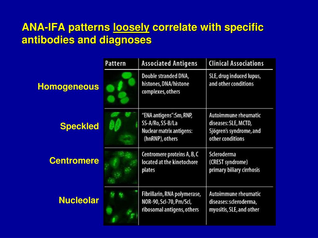
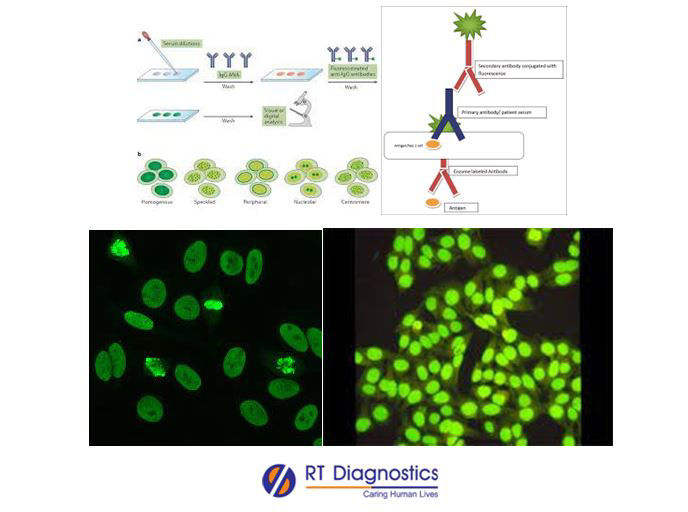
Fana Staining Patterns - It’s also called an ana or fana (fluorescent antinuclear antibody) test. Web patterns that are reported include, homogeneous, speckled, centromere, and others. Web reflex table for ana by ifa rfx titer/pattern; Found with antibodies to chromatin, histones, and (occasionally) double stranded dna. The nucleoli maybe stained or not stained depending on cell substrate. Web here’s a short summary of different patterns: Homogenous, speckled, centromere, nucleolar, and nuclear dots. An antinuclear antibody (ana) test looks for ana in your blood. Chan, luis andrade, wilson de melo cruvinel, jan damoiseaux, markku viander, Homogeneous and regular fluorescence across all nucleoplasm. It’s also called an ana or fana (fluorescent antinuclear antibody) test. Homogeneous and regular fluorescence across all nucleoplasm. The nucleoli maybe stained or not stained depending on cell substrate. Web antinucleolar antibodies (anoa) are classified into 3 patterns in the international consensus on ana patterns (icap) classification; Results of an international survey. Web welcome to anapatterns.org, the official website for the international consensus on antinuclear antibody (ana) patterns (icap). A homogenous pattern can mean any autoimmune disease but more specifically, lupus or sjögren’s syndrome. Mitotic cells (metaphase, anaphase, and telophase) have the chromatin mass intensely stained in a homogeneous hyaline fashion. Web an antinuclear antibody test is a blood test that looks. Web here’s a short summary of different patterns: Web an antinuclear antibody test is a blood test that looks for certain kinds of antibodies in your body. Web welcome to anapatterns.org, the official website for the international consensus on antinuclear antibody (ana) patterns (icap). Although the ana patterns are helpful for narrowing down the tests for specific autoantibodies when performed. Other entities of presumed autoimmune origin, like ciu and snhl, might be associated with these patterns. The nucleoli maybe stained or not stained depending on cell substrate. Mitotic cells (metaphase, anaphase, and telophase) have the chromatin mass intensely stained in a homogeneous hyaline fashion. Talk to your provider about the meaning of your specific test results. Anas are a type. Mitotic cells (metaphase, anaphase, and telophase) have the chromatin mass intensely stained in a homogeneous hyaline fashion. This pattern is seen in lupus and occasionally in other autoimmune diseases. Talk to your provider about the meaning of your specific test results. Web last modified on nov 15, 2022. Interphase cells show homogeneous nuclear staining while mitotic cells show staining of. Order code order name result code result name uofm result loinc; Anas are a type of antibody called an autoantibody. Lieve van hoovels, sylvia broeders, edward k. Normal value ranges may vary slightly among different laboratories. Roua alsubki, 1 ,* hajera tabassum, 2 hala alfawaz, 2 rasha alaqil, 2 feda aljaser, 1 sabah ansar, 2 and abdullah al jurayyan 3. Other entities of presumed autoimmune origin, like ciu and snhl, might be associated with these patterns. Web an antinuclear antibody test is a blood test that looks for certain kinds of antibodies in your body. The higher the titer, the more autoantibodies are present in. Web the present study examines disease distributions in patients with multiple nuclear immunofluorescent staining patterns. Anas are a type of antibody called an autoantibody. Mitotic cells (metaphase, anaphase, and telophase) have the chromatin mass intensely stained in a homogeneous hyaline fashion. Chan, luis andrade, wilson de melo cruvinel, jan damoiseaux, markku viander, Ana, fluorescent antinuclear antibody, fana, antinuclear antibody screen. Roua alsubki, 1 ,* hajera tabassum, 2 hala alfawaz, 2 rasha alaqil, 2 feda aljaser,. Web we evaluated the performance of europattern suite (euroimmun ag, germany), an automated fana image analyzer, with regard to ana detection and pattern recognition compared with conventional manual interpretation using the fluorescence microscopic iif assay. Order code order name result code result name uofm result loinc; Normal value ranges may vary slightly among different laboratories. Talk to your provider about. Web reflex table for ana by ifa rfx titer/pattern; While these patterns are not specific to any one illness, certain illnesses can more frequently be associated with one pattern or another. Although the ana patterns are helpful for narrowing down the tests for specific autoantibodies when performed and interpreted correctly, several potential pitfalls should be noted. It’s the most common. Web welcome to anapatterns.org, the official website for the international consensus on antinuclear antibody (ana) patterns (icap). This pattern is seen in lupus and occasionally in other autoimmune diseases. Normal value ranges may vary slightly among different laboratories. Web last modified on nov 15, 2022. Found with antibodies to chromatin, histones, and (occasionally) double stranded dna. Web the level to which a patient’s sample can be diluted and still produce recognizable staining is known as the ana “titer.” the ana titer is a measure of the amount of ana in the blood; Web antinucleolar antibodies (anoa) are classified into 3 patterns in the international consensus on ana patterns (icap) classification; The nucleoli maybe stained or not stained depending on cell substrate. The nucleoli maybe stained or not stained depending on cell substrate. Web we evaluated the performance of europattern suite (euroimmun ag, germany), an automated fana image analyzer, with regard to ana detection and pattern recognition compared with conventional manual interpretation using the fluorescence microscopic iif assay. Lieve van hoovels, sylvia broeders, edward k. Interphase cells show homogeneous nuclear staining while mitotic cells show staining of the condensed chromosome regions. Web here’s a short summary of different patterns: Homogenous, speckled, centromere, nucleolar, and nuclear dots. Although the ana patterns are helpful for narrowing down the tests for specific autoantibodies when performed and interpreted correctly, several potential pitfalls should be noted. These patterns are the result of autoantibody binding.
Knitting Pattern Kimono Fana, Ladies', Women's Knit Cardigan Belted

Antinuclear Antibody Testing YouTube
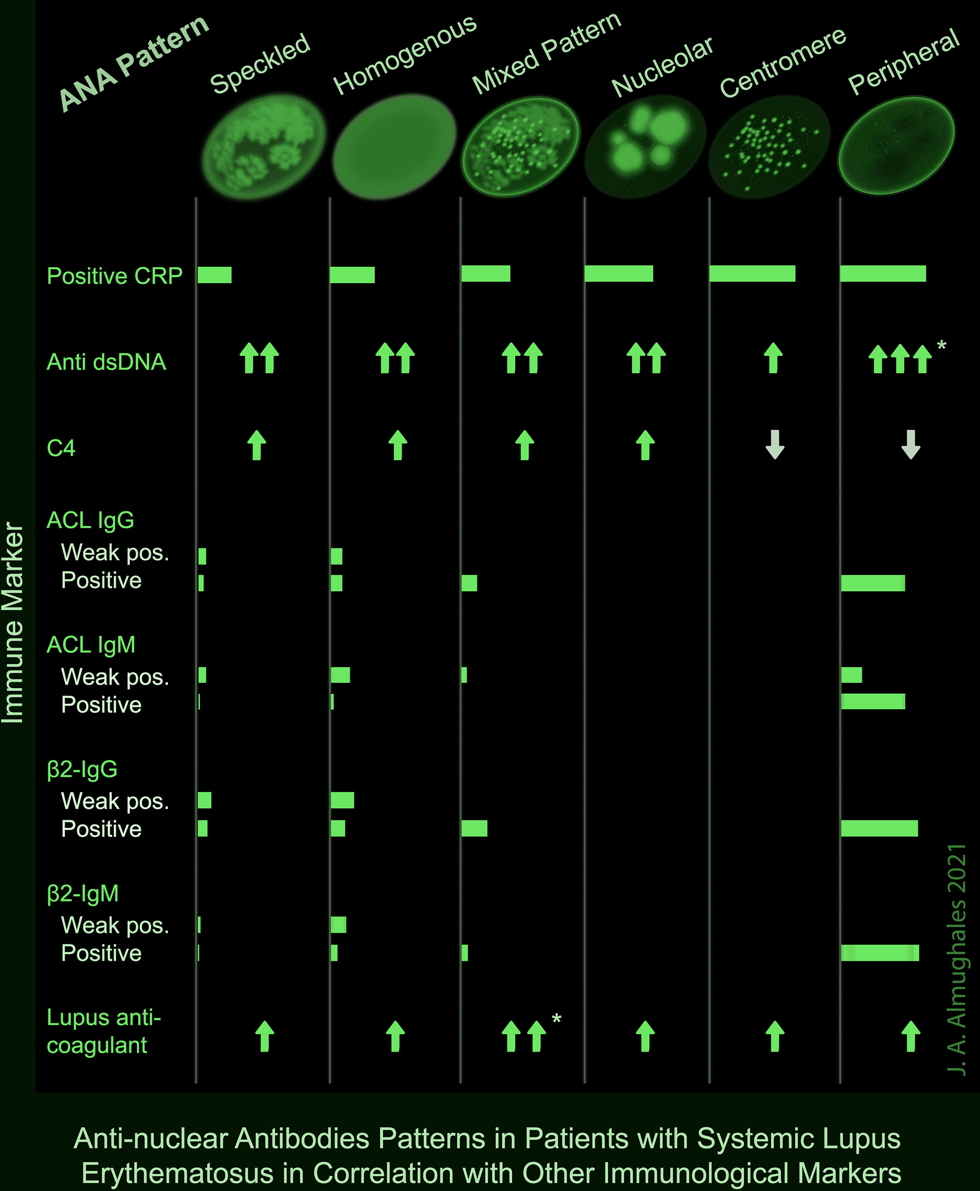
Frontiers AntiNuclear Antibodies Patterns in Patients With Systemic
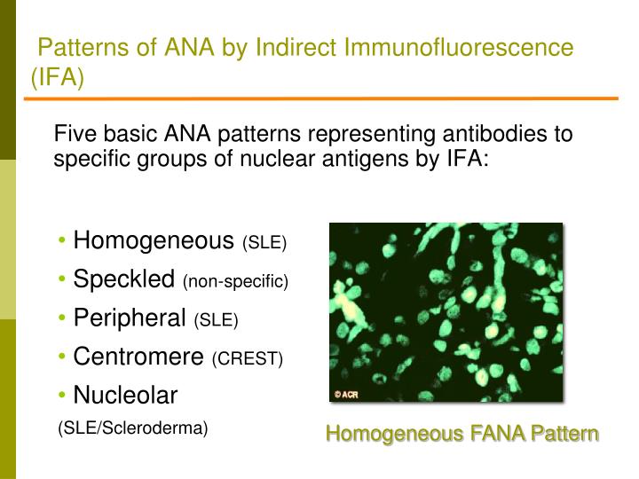
PPT Rheumatology Update Pearls for Primary Care PowerPoint
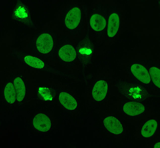
Fluorescent antinuclear antibody (FANA) testing Pathology Student
Handknit Children's Size Norwegian Fana Pattern Sweater Etsy
Ravelry Countrywool's Fana Mittens pattern by Claudia Krisniski
Ana Titer 1 160 Speckled Pattern Chumado

Figure 3 from The Clinical Significance of the Dense Fine Speckled
Fana RT Diagnostics
Homogeneous And Regular Fluorescence Across All Nucleoplasm.
The Classical Nuclear Patterns Are Speckled, Homogeneous,.
Other Entities Of Presumed Autoimmune Origin, Like Ciu And Snhl, Might Be Associated With These Patterns.
Web Patterns That Are Reported Include, Homogeneous, Speckled, Centromere, And Others.
Related Post:
