Ekg Pulmonary Disease Pattern
Ekg Pulmonary Disease Pattern - Web electrocardiography (ecg) is a useful adjunct to other pulmonary tests because it provides information about the right side of the heart and therefore pulmonary disorders such as chronic pulmonary hypertension and pulmonary embolism. Web background—chronic cor pulmonale (ccp) is a strong predictor of death in chronic obstructive pulmonary disease (copd). Ecg changes commonly associated with pulmonary diseases such as copd. Web objective patients with chronic obstructive pulmonary disease (copd) often have abnormal ecgs. This pattern is characterized by a large s wave in lead i, a q wave in lead iii, and an inverted t wave in lead iii. Web the principal electrocardiogram (ecg) changes associated with ventricular hypertrophy are increases in qrs amplitude and duration, changes in instantaneous and mean qrs vectors, abnormalities in the st segment and t waves, and abnormalities in the p wave. Web electrocardiography (ecg) is a useful adjunct to other pulmonary tests because it provides information about the right side of the heart and therefore pulmonary disorders such as chronic pulmonary hypertension and pulmonary embolism. Web patients with chronic obstructive pulmonary disease (copd) often have abnormal ecgs. Web ecg changes occur in chronic obstructive pulmonary disease (copd) due to: The distances between these points and. Patients presenting with chest pain, these ekg patterns, and troponin elevation are often misdiagnosed with mi. Ecgs were interpreted blindly in 63 patients with severe copd (group 1) versus 83 patients with mild or moderate copd (group 2). Web patients with chronic obstructive pulmonary disease (copd) often have abnormal ecgs. Our aim was to separate the effects on ecg by. Web ecg abnormalities are common in patients with pulmonary embolism, with the most frequent being sinus tachycardia, right ventricular strain, and the classic s1q3t3 pattern. Our aim was to separate the effects on ecg by airway obstruction, emphysema and right ventricular (rv) afterload in patients with copd. Ecg changes commonly associated with pulmonary diseases such as copd. Web background—chronic cor. Our aim was to separate the effects on ecg by airway obstruction, emphysema and right ventricular (rv) afterload in patients with copd. Web sinus rhythm (which is the normal rhythm) has the following characteristics: Web the ecg changes associated with acute pulmonary embolism may be seen in any condition that causes acute pulmonary hypertension, including hypoxia causing pulmonary hypoxic vasoconstriction.. The presence of hyperexpanded emphysematous lungs within the chest; Web based on the low voltage in leads v 1, v 2, v 3, the rightward frontal plane axis, incomplete right bundle branch block (rbbb), and persistent precordial s waves, the computer interpreted the overall pattern as consistent with pulmonary disease. Web this post describes two ekg patterns of pe which. Web electrocardiography (ecg) is a useful adjunct to other pulmonary tests because it provides information about the right side of the heart and therefore pulmonary disorders such as chronic pulmonary hypertension and pulmonary embolism. Ecg changes commonly associated with pulmonary diseases such as copd. Patients presenting with chest pain, these ekg patterns, and troponin elevation are often misdiagnosed with mi.. Our aim was to separate the effects on ecg by airway obstruction, emphysema and right ventricular (rv) afterload in patients with copd. Again, this indicates significant right ventricular strain. Web sinus rhythm (which is the normal rhythm) has the following characteristics: Common ecg patterns seen in ph include right atrial abnormalities, right axis deviation, right ventricular hypertrophy with strain pattern.. Patients presenting with chest pain, these ekg patterns, and troponin elevation are often misdiagnosed with mi. Web ecg changes occur in chronic obstructive pulmonary disease (copd) due to: •right axis deviation or vertical axis of the qrs complex. Common ecg patterns seen in ph include right atrial abnormalities, right axis deviation, right ventricular hypertrophy with strain pattern. Ecgs were interpreted. Web the ecg changes associated with acute pulmonary embolism may be seen in any condition that causes acute pulmonary hypertension, including hypoxia causing pulmonary hypoxic vasoconstriction. Again, this indicates significant right ventricular strain. The aims of this study were to assess the prognostic role of individual ecg signs of ccp and of the interaction The presence of hyperexpanded emphysematous lungs. However, its prognostic role in patients with ph remains uncertain. We used different criteria for the s1s2s3 ecg pattern. Web electrocardiography (ecg) is a useful adjunct to other pulmonary tests because it provides information about the right side of the heart and therefore pulmonary disorders such as chronic pulmonary hypertension and pulmonary embolism. Web chronic obstructive pulmonary diseases (copd), a. •right axis deviation or vertical axis of the qrs complex. Web electrocardiography (ecg) is a useful adjunct to other pulmonary tests because it provides information about the right side of the heart and therefore pulmonary disorders such as chronic pulmonary hypertension and pulmonary embolism. Ecgs were interpreted blindly in 63 patients with severe copd (group 1) versus 83 patients with. Web ecg abnormalities are common in patients with pulmonary embolism, with the most frequent being sinus tachycardia, right ventricular strain, and the classic s1q3t3 pattern. Our aim was to separate the effects on ecg by airway obstruction, emphysema and right ventricular (rv) afterload in patients with copd. Web electrocardiography (ecg) is a useful adjunct to other pulmonary tests because it provides information about the right side of the heart and therefore pulmonary disorders such as chronic pulmonary hypertension and pulmonary embolism. This pattern is characterized by a large s wave in lead i, a q wave in lead iii, and an inverted t wave in lead iii. The prevalence of some electrocardiographic (ecg) abnormalities in severe versus mild or moderate chronic obstructive pulmonary disease (copd) has been reported. Web the principal electrocardiogram (ecg) changes associated with ventricular hypertrophy are increases in qrs amplitude and duration, changes in instantaneous and mean qrs vectors, abnormalities in the st segment and t waves, and abnormalities in the p wave. Again, this indicates significant right ventricular strain. Web electrocardiography (ecg) is a useful adjunct to other pulmonary tests because it provides information about the right side of the heart and therefore pulmonary disorders such as chronic pulmonary hypertension and pulmonary embolism. Web electrocardiography (ecg) is a useful adjunct to other pulmonary tests because it provides information about the right side of the heart and therefore pulmonary disorders such as chronic pulmonary hypertension and pulmonary embolism. Ecg changes commonly associated with pulmonary diseases such as copd. Ecgs were interpreted blindly in 63 patients with severe copd (group 1) versus 83 patients with mild or moderate copd (group 2). Common ecg patterns seen in ph include right atrial abnormalities, right axis deviation, right ventricular hypertrophy with strain pattern. However, its prognostic role in patients with ph remains uncertain. Web sinus rhythm (which is the normal rhythm) has the following characteristics: Web the ecg changes associated with acute pulmonary embolism may be seen in any condition that causes acute pulmonary hypertension, including hypoxia causing pulmonary hypoxic vasoconstriction. Web patients with chronic obstructive pulmonary disease (copd) often have abnormal ecgs.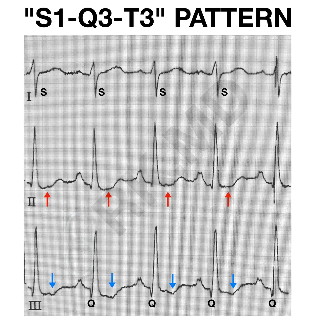
S1Q3T3 EKG Pattern RK.MD
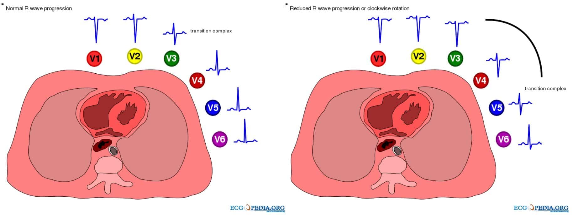
ECG in Chronic Obstructive Pulmonary Disease • LITFL • ECG Library

S1Q3T3 EKG Classic Pattern in Pulmonary Embolism (Example). Pulmonary

Schematic representations of ECG patterns. Normal ECG pattern with
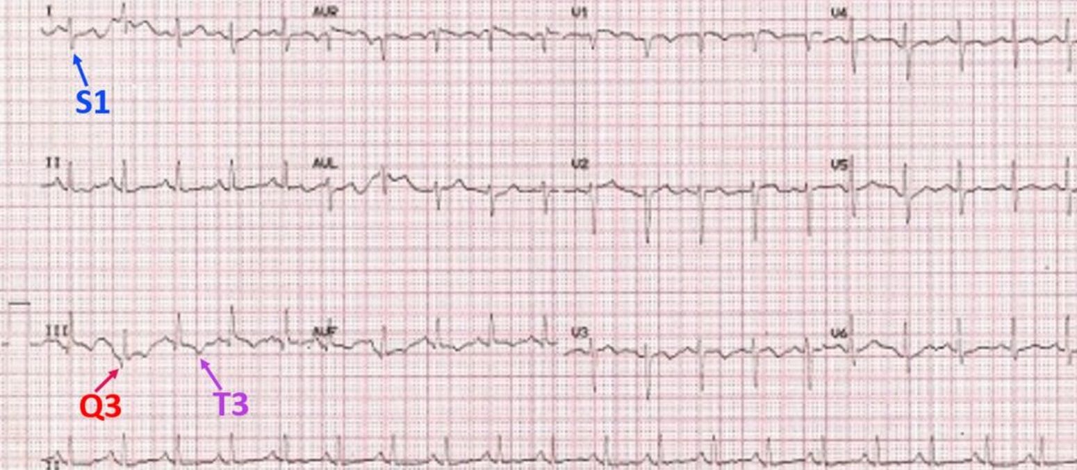
S1Q3T3 pattern on ECG in pulmonary embolism All About Cardiovascular
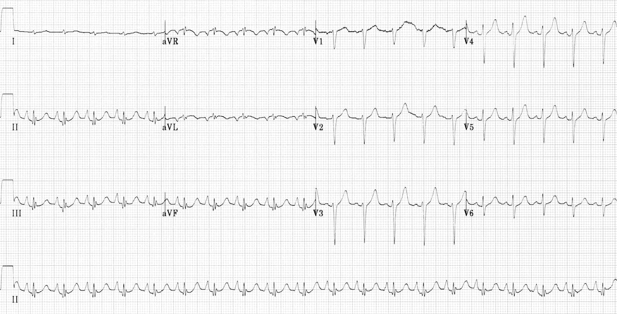
ECG in Chronic Obstructive Pulmonary Disease • LITFL • ECG Library
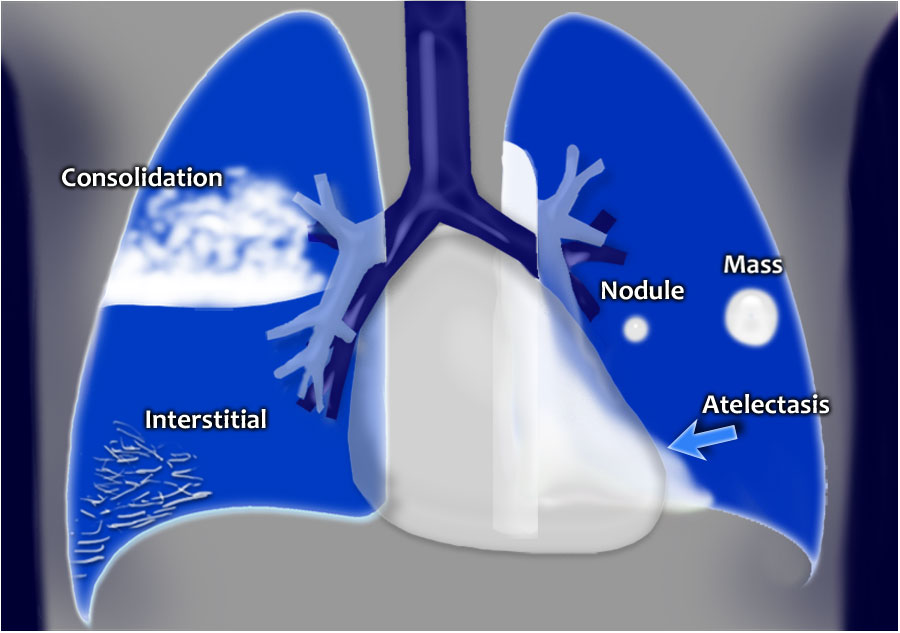
Chest XRay Lung disease FourPattern Approach NCLEX Quiz

S1Q3T3 on ECG in a patient with Acute Pulmonary Embolism GrepMed

ECG Changes in Pulmonary Embolism New Health Advisor

The ECG's of Pulmonary Embolism Resus
Ecg Findings Often Suggest Right Ventricular Pressure Overload Or Strain.
What Else Should Be Added To Your Interpretation?
Web An Ecg Readout Represents The Pattern Of Electrical Activity In The Heart As A Line Of Waves.
Web Ecg Changes Occur In Chronic Obstructive Pulmonary Disease (Copd) Due To:
Related Post: