Centromere Pattern
Centromere Pattern - Scleroderma 70 antibody aka topoisomerase 1. The highly repetitive and epigenetic nature of centromeres were documented during the past half century. A centromere pattern may indicate scleroderma. During mitosis, spindle fibers attach to the centromere via the kinetochore. T is true that a centromere ana pattern is associated with crest. The centromere is the point on a chromosome where mitotic spindle fibers attach to pull sister chromatids apart during cell division. Web a speckled pattern may indicate various diseases, including lupus and sjögren’s syndrome. Autoantibodies to centromere antigens are found in 22% of patients with progressive systemic sclerosis (pss, or diffuse scleroderma) and in 90% of patients with the subset of scleroderma known as the crest syndrome (calcinosis, raynauds, esophageal. Web the centromere is the region of the chromosome that directs its segregation in mitosis and meiosis. Although the functional importance of the centromere has been appreciated for more than 130. The centromere appears as a constricted region of a chromosome and plays a key role in helping the cell divide up its dna during division (mitosis and meiosis). Autoantibodies to centromere antigens are found in 22% of patients with progressive systemic sclerosis (pss, or diffuse scleroderma) and in 90% of patients with the subset of scleroderma known as the crest. Positive antinuclear antibody test with centromere pattern. Web the centromere links a pair of sister chromatids together during cell division. Screening for this antibody should be conducted in all patients with raynaud's phenomenon, primary biliary cirrhosis, and scleroderma. However, characteristics of aca in comparison with the other ana patterns and clinical. Web a centromere pattern is highly associated with ssc. This constricted region of chromosome connects the sister chromatids, creating a short arm (p) and a long arm (q) on the chromatids. Specifically, it is the region where the cell’s spindle fibers attach. A centromere staining pattern means the ana staining is present along the chromosomes. Positive antinuclear antibody test with centromere pattern. The highly repetitive and epigenetic nature of. A nucleolar staining pattern means ana staining is present around the. A centromere staining pattern means the ana staining is present along the chromosomes. The centromere is the genetic locus that specifies the site of kinetochore assembly, where the chromosome will attach to the kinetochore microtubule. Screening for this antibody should be conducted in all patients with raynaud's phenomenon, primary. The centromere is the genetic locus that specifies the site of kinetochore assembly, where the chromosome will attach to the kinetochore microtubule. Web in this review, we synthesize the research on the central features of centromere identity, the molecular basis for centromere propagation to the chromosomes of daughter cells and gametes, and the mechanisms by which the centromere recruits the. Unusual staining patterns in nuclei, such as those for the nuclear mitotic apparatus (numa) [ 3 ] ( fig. Specifically, it is the region where the cell’s spindle fibers attach. Unique chromatin structures that drive chromosome segregation. Screening for this antibody should be conducted in all patients with raynaud's phenomenon, primary biliary cirrhosis, and scleroderma. During mitosis, spindle fibers attach. Seen in people with lupus or diffuse scleroderma. The centromere appears as a constricted region of a chromosome and plays a key role in helping the cell divide up its dna during division (mitosis and meiosis). Web the centromere is the region of the chromosome that directs its segregation in mitosis and meiosis. Scleroderma 70 antibody aka topoisomerase 1. Unusual. The highly repetitive and epigenetic nature of centromeres were documented during the past half century. Although the functional importance of the centromere has been appreciated for more than 130. During mitosis, spindle fibers attach to the centromere via the kinetochore. Unique chromatin structures that drive chromosome segregation. Web in this review, we synthesize the research on the central features of. Web centromeres are the chromosomal domains required to ensure faithful transmission of the genome during cell division. However, characteristics of aca in comparison with the other ana patterns and clinical. They have a central role in preventing aneuploidy, by orchestrating the. Web positive antinuclear antibody test with nucleolar pattern. 1d ), may not be reported depending on the. A nucleolar staining pattern means ana staining is present around the. Web a centromere pattern is highly associated with ssc 5,8. Although the functional importance of the centromere has been appreciated for more than 130. Web centromeres are the chromosomal domains required to ensure faithful transmission of the genome during cell division. The centromere is the genetic locus that specifies. Web a centromere pattern is usually reported as distinct pattern, but they can also be termed discrete speckled nuclear staining patterns (fig. The highly repetitive and epigenetic nature of centromeres were documented during the past half century. This constricted region of chromosome connects the sister chromatids, creating a short arm (p) and a long arm (q) on the chromatids. A centromere pattern may indicate scleroderma. The centromere is the point on a chromosome where mitotic spindle fibers attach to pull sister chromatids apart during cell division. (1,2) aca has a broad specificity for the. Web a speckled pattern may indicate various diseases, including lupus and sjögren’s syndrome. Web in this review, we synthesize the research on the central features of centromere identity, the molecular basis for centromere propagation to the chromosomes of daughter cells and gametes, and the mechanisms by which the centromere recruits the kinetochore to establish a connection to spindle microtubules. A centromere staining pattern means the ana staining is present along the chromosomes. Ana patterns are frequently interpreted incorrectly and thus i would question the result. Seen in people with limited scleroderma. Web the anticentromere antibody is therefore a useful prognostic indicator in patients with early scleroderma, as it may help to predict what pattern of scleroderma will evolve. However, characteristics of aca in comparison with the other ana patterns and clinical. Unique chromatin structures that drive chromosome segregation. During mitosis, spindle fibers attach to the centromere via the kinetochore. T is true that a centromere ana pattern is associated with crest.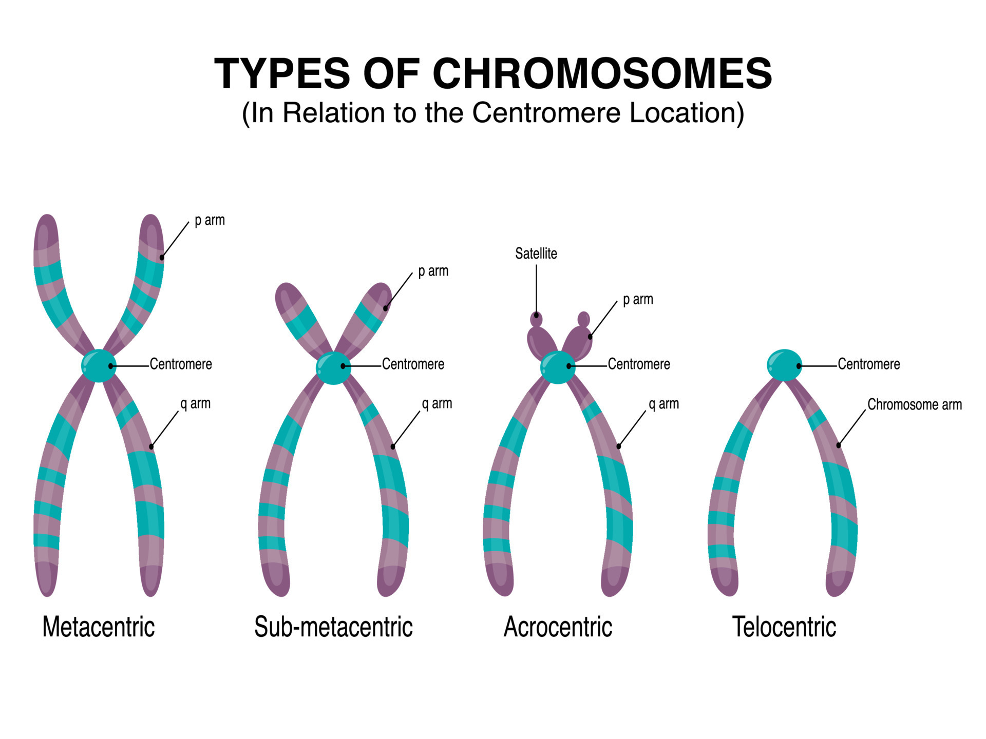
Types of Chromosomes in relation to the centromere location 7508619
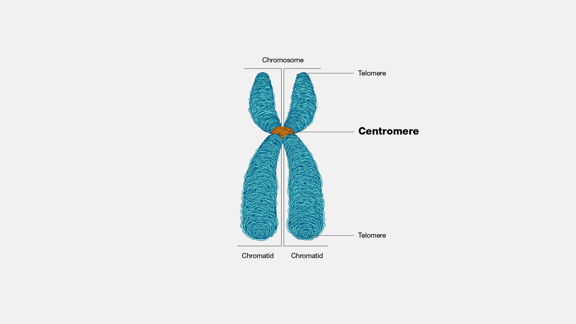
Centromere

Centromere staining pattern in indirect fluorescence Download

Figure 1 Determining centromere identity cyclical stories and
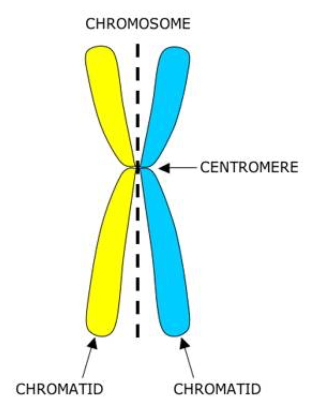
Centromere Structure
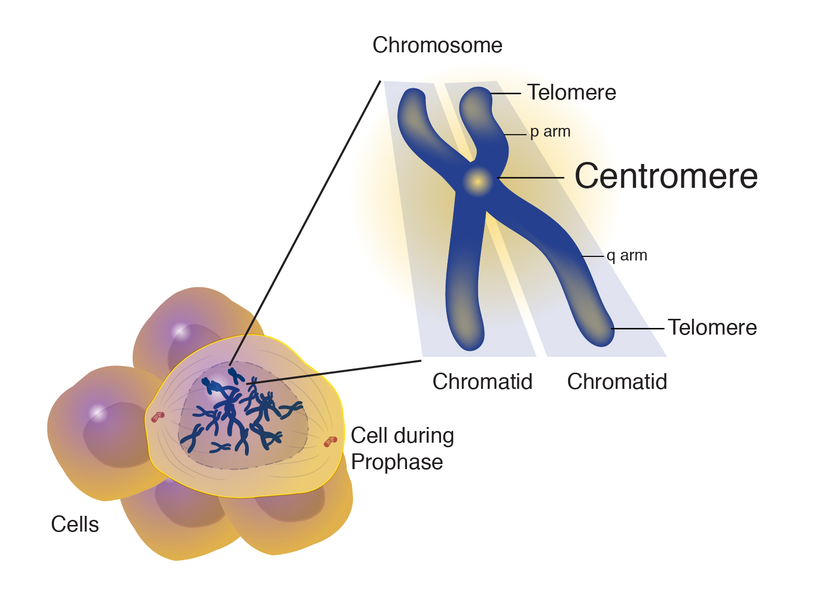
Centromere
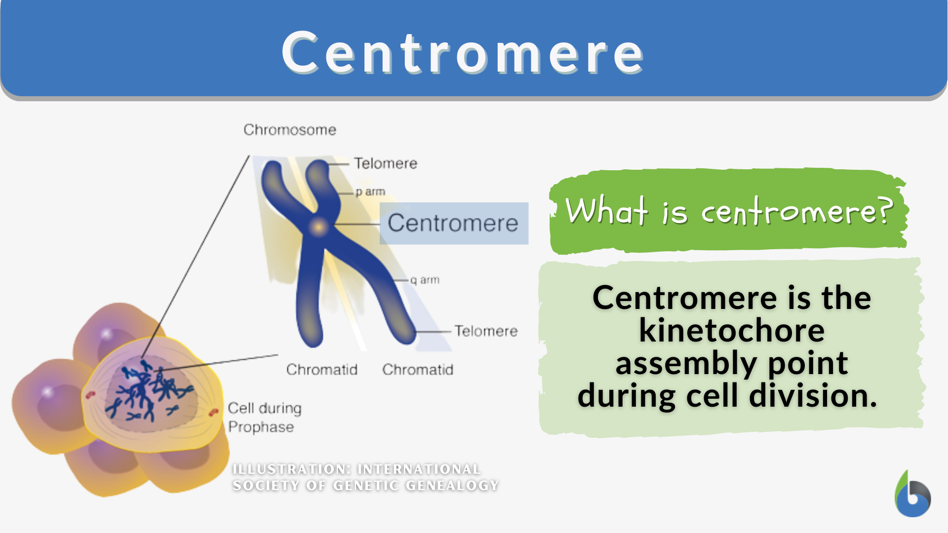
Centromere Definition and Examples Biology Online Dictionary

Centromere Definition, Structure, Position, Types, Functions
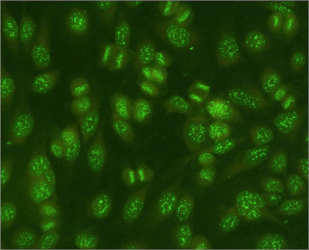
Antinuclear antibodies (ANA) positive control centromere pattern
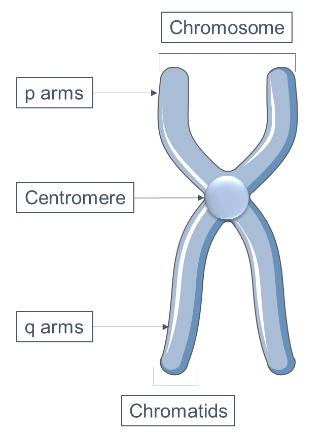
Centromere Structure
Although The Functional Importance Of The Centromere Has Been Appreciated For More Than 130.
Screening For This Antibody Should Be Conducted In All Patients With Raynaud's Phenomenon, Primary Biliary Cirrhosis, And Scleroderma.
Autoantibodies To Centromere Antigens Are Found In 22% Of Patients With Progressive Systemic Sclerosis (Pss, Or Diffuse Scleroderma) And In 90% Of Patients With The Subset Of Scleroderma Known As The Crest Syndrome (Calcinosis, Raynauds, Esophageal.
Web The Centromere Links A Pair Of Sister Chromatids Together During Cell Division.
Related Post: