Brugada Ecg Patterns
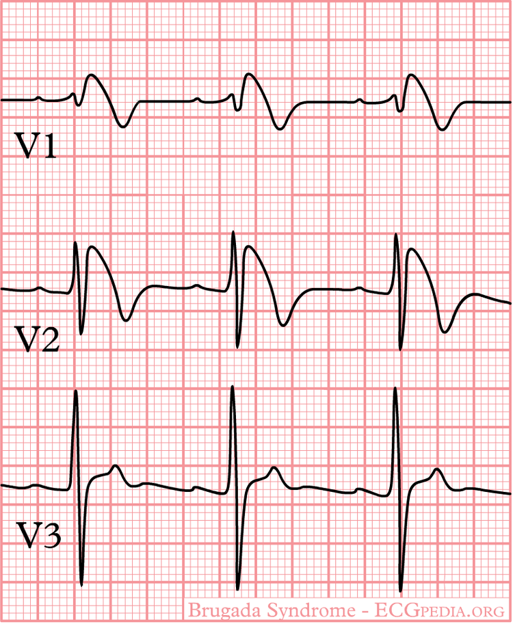
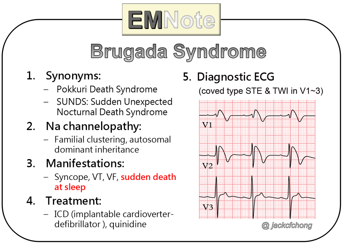
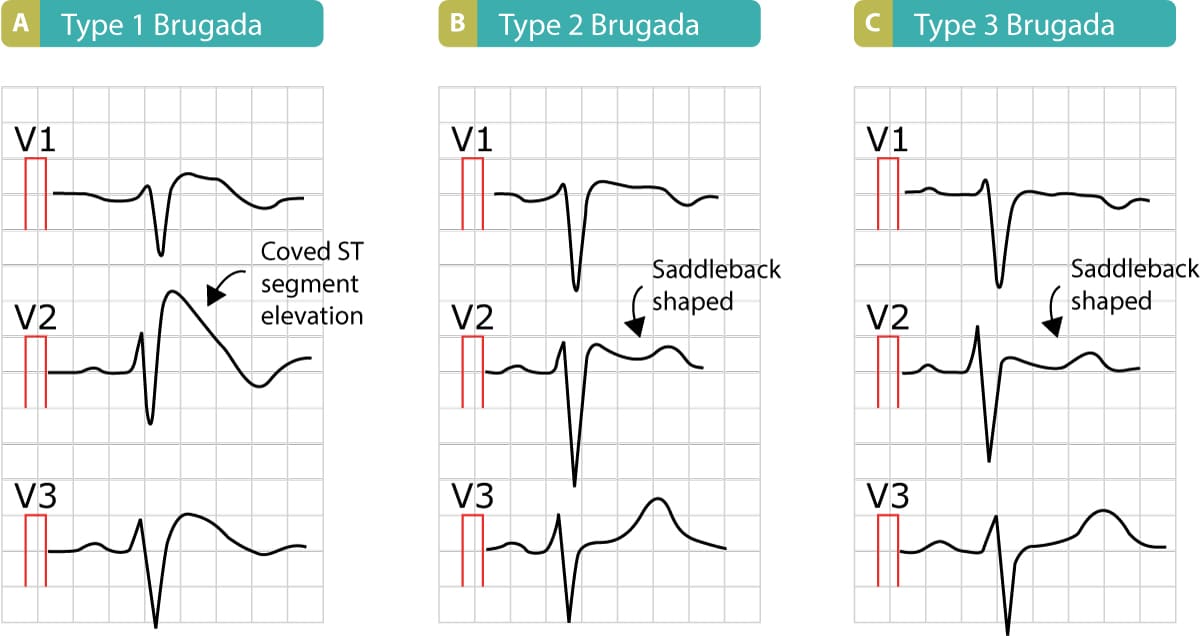
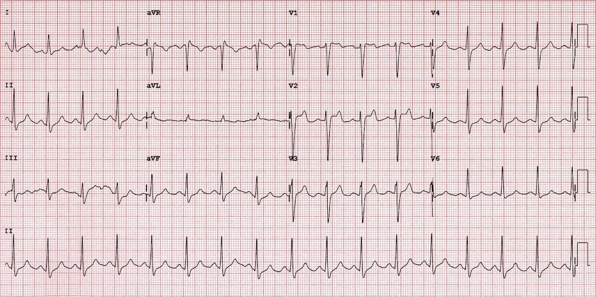
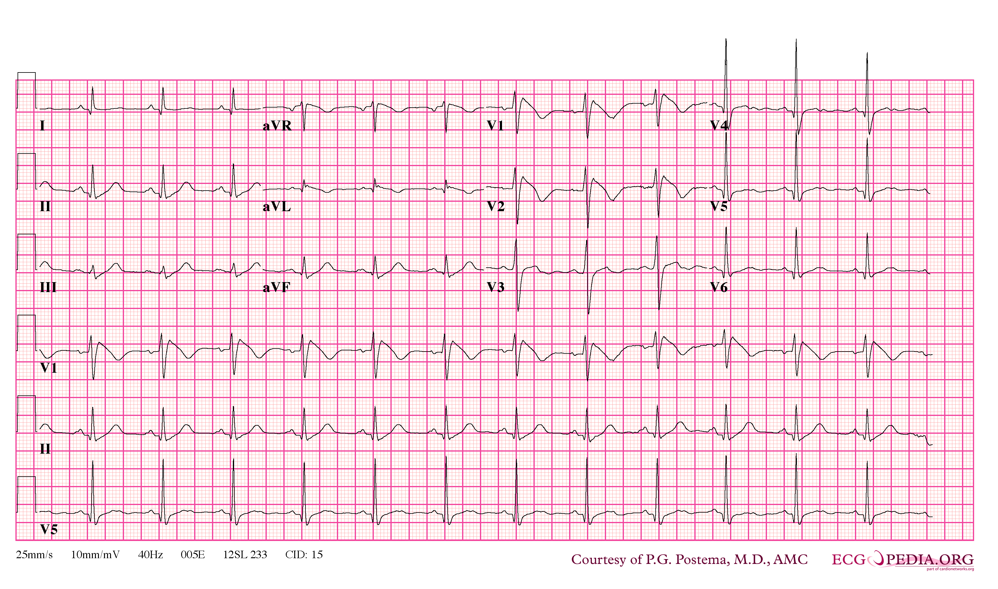
Brugada Ecg Patterns - It increases the risk of abnormal heart rhythms and sudden cardiac death. The mean age of sudden death is 41, with the age at diagnosis ranging from 2 days to 84 years. Web brugada syndrome (brs) is an inherited sodium, calcium, or potassium channelopathy associated with an increased risk of ventricular fibrillation (vf) and sudden cardiac death. Their father showed a spontaneous type 1 brugada electrocardiogram pattern. Web the brugada ecg pattern is modulated by genetic mutations and pharmacological agents that alter the function of ion channels active during the early phases of the action potential. Web the brugada pattern, which occurs in asymptomatic individuals without any other clinical criteria, has an estimated prevalence of between 0.1% and 1%. The type 1 brugada ecg pattern has prominent st elevation in v1 and v2 (sometimes involving v3) that causes the qrs complex in these leads to resemble right bundle branch block. Web sudden cardiac death (scd) is responsible for ≈50% of all cardiovascular deaths and is the first clinical manifestation of a previously subclinical cardiac disease in ≈50% of the cases. Three repolarisation patterns are associated with bs4 (figure 1) when found in more than one right precordial leads (v1 to v3): Journal of the american college of cardiology. Brugada syndrome signs and symptoms are similar to those of some other heart rhythm disorders. Brugada syndrome is a rare condition that causes an abnormal heart rhythm in your heart's lower chambers (ventricles). Their father showed a spontaneous type 1 brugada electrocardiogram pattern. The abnormal heart rhythms seen in those with brugada syndrome often occur at. This irregular heartbeat can. Brugada syndrome is an inherited channelopathy causing an increased risk of ventricular tachycardia (vt) and ventricular fibrillation (vf) leading to syncope and sudden death. Web three ecg repolarization patterns in the right precordial leads are recognized in the diagnosis of brugada syndrome. Furthermore, the proband’s genetic testing. Brugada syndrome signs and symptoms are similar to those of some other heart. It increases the risk of abnormal heart rhythms and sudden cardiac death. Web the brugada syndrome may present with three different ecg patterns, referred to as type 1, type 2, and type 2 brugada syndrome ecg. The mean age of sudden death is 41, with the age at diagnosis ranging from 2 days to 84 years. Patients have abnormal findings. (see also overview of arrhythmias and overview of channelopathies.) Furthermore, signs of right ventricular structural abnormalities are often found in patients with brugada syndrome. Web the brugada syndrome may present with three different ecg patterns, referred to as type 1, type 2, and type 2 brugada syndrome ecg. Web brugada syndrome is an ecg abnormality with a high incidence of. Web the brugada pattern, which occurs in asymptomatic individuals without any other clinical criteria, has an estimated prevalence of between 0.1% and 1%. Correct recognition of the diagnostic brugada syndrome ecg pattern. Looking for the signs of brugada. The most typical, and diagnostic, is type 1 brugada syndrome. Web a major sign of brugada syndrome is an irregular result on. Correct recognition of the diagnostic brugada syndrome ecg pattern. Journal of the american college of cardiology. The type 1 brugada ecg pattern has prominent st elevation in v1 and v2 (sometimes involving v3) that causes the qrs complex in these leads to resemble right bundle branch block. Web brugada syndrome is an ecg abnormality with a high incidence of sudden. Brugada syndrome is an ecg abnormality with a high incidence of sudden death in patients with structurally normal hearts. When to see a doctor. A heterozygous scn5a p.r893c variant was found by genetic testing in the proband’s father and sister. Type 1 brugada ecg pattern. This irregular heartbeat can cause fainting (syncope) and lead to sudden cardiac death (scd). A heterozygous scn5a p.r893c variant was found by genetic testing in the proband’s father and sister. Brugada syndrome is a rare condition that causes an abnormal heart rhythm in your heart's lower chambers (ventricles). The mean age of sudden death is 41, with the age at diagnosis ranging from 2 days to 84 years. Web initial diagnosis of brugada syndrome. Web brugada syndrome (brs) is an inherited sodium, calcium, or potassium channelopathy associated with an increased risk of ventricular fibrillation (vf) and sudden cardiac death. Brugada syndrome poses significant challenges in terms of risk stratification and management, particularly for asymptomatic patients who comprise the majority of individuals exhibiting brugada ecg pattern (brecg). Web brugada syndrome ( brs) is a genetic. The abnormal heart rhythms seen in those with brugada syndrome often occur at. Web five years later, his sister showed brugada electrocardiogram pattern while febrile from kawasaki disease. This often happens while you’re at rest or asleep. Brugada syndrome signs and symptoms are similar to those of some other heart rhythm disorders. Brugada syndrome is an ecg abnormality with a. Web five years later, his sister showed brugada electrocardiogram pattern while febrile from kawasaki disease. Correct recognition of the diagnostic brugada syndrome ecg pattern. Brugada syndrome is an inherited channelopathy causing an increased risk of ventricular tachycardia (vt) and ventricular fibrillation (vf) leading to syncope and sudden death. Web the brugada syndrome may present with three different ecg patterns, referred to as type 1, type 2, and type 2 brugada syndrome ecg. 1) there is one true diagnostic of the brugada pattern; Brugada syndrome is an ecg abnormality with a high incidence of sudden death in patients with structurally normal hearts. This irregular heartbeat can cause fainting (syncope) and lead to sudden cardiac death (scd). Web brugada syndrome (brs) is an inherited sodium, calcium, or potassium channelopathy associated with an increased risk of ventricular fibrillation (vf) and sudden cardiac death. Web the brugada pattern, which occurs in asymptomatic individuals without any other clinical criteria, has an estimated prevalence of between 0.1% and 1%. Web initial diagnosis of brugada syndrome is based on a characteristic ecg pattern, the type 1 brugada ecg pattern (see figure type 1 brugada ecg pattern). Looking for the signs of brugada. A heterozygous scn5a p.r893c variant was found by genetic testing in the proband’s father and sister. Web three ecg repolarization patterns in the right precordial leads are recognized in the diagnosis of brugada syndrome. This often happens while you’re at rest or asleep. The most typical, and diagnostic, is type 1 brugada syndrome. The type i ecg is characterized by a j elevation >=2 mm (0.2 mv) a coved type st segment followed by a negative t wave (see figure).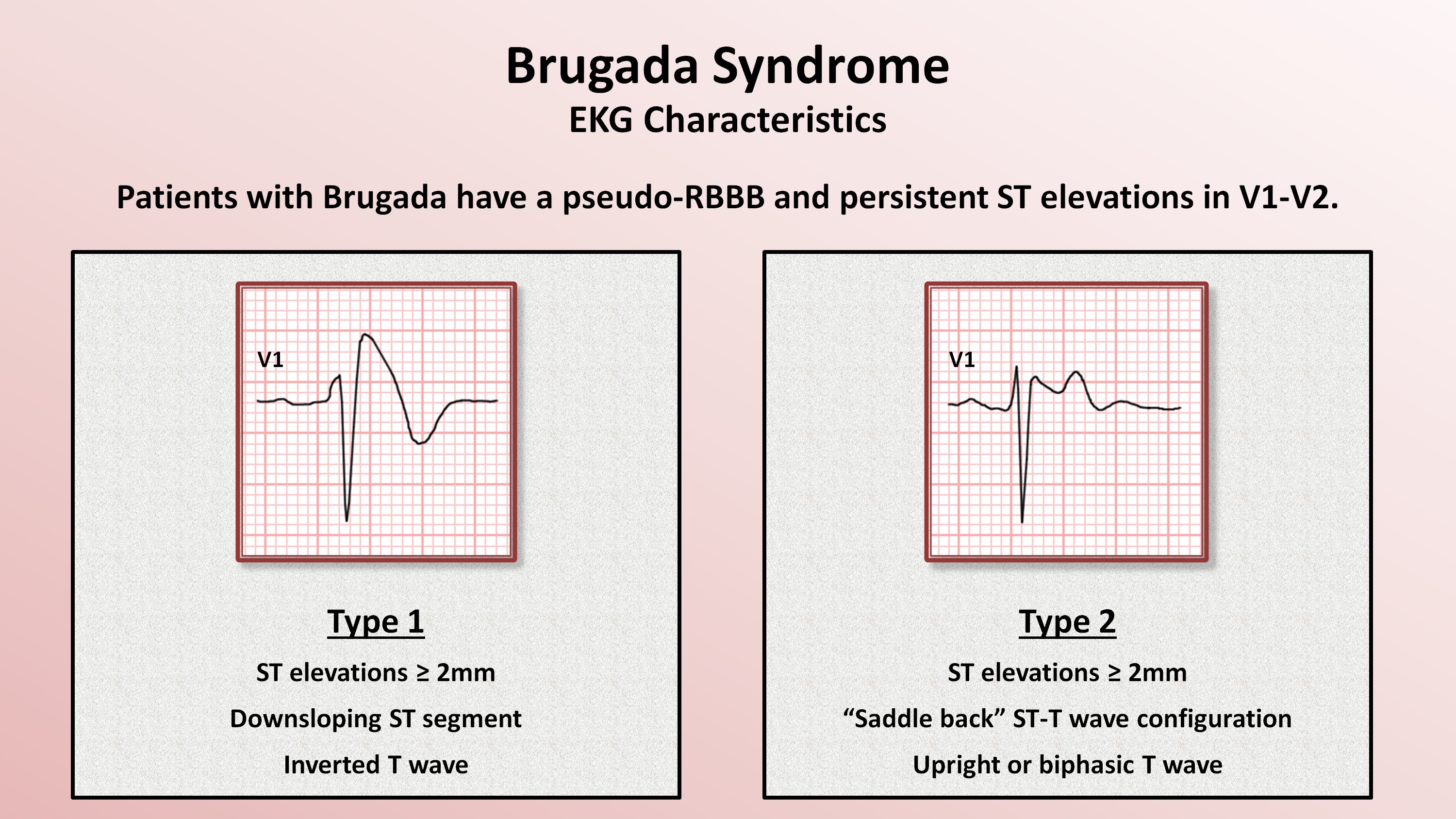
Brugada Syndrome

Brugada Syndrome Causes, ECG, Symptoms, Treatment
Brugada Syndrome
Brugada syndrome,what to know?
Brugada syndrome ECG, clinical features and management ECG learning
Brugada Syndrome • LITFL • ECG Library Diagnosis
FileBrugada syndrome type1 example4.png ECGpedia

FileBrugada syndrome type1 example1.png ECGpedia
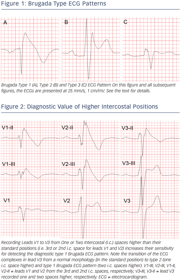
Figure 1 Brugada Type ECG Patterns Radcliffe Vascular

Brugada Syndrome Causes, ECG, Symptoms, Treatment
Web This Pattern In The Presence Of Documented Ventricular Arrhythmias Or Its Symptoms (Syncope, Seizure) Or Significant Family For Sudden Cardiac Death Or Abovementioned Ecg Changes Is Called Brugada Syndrome.
Web Brugada Syndrome ( Brs) Is A Genetic Disorder In Which The Electrical Activity Of The Heart Is Abnormal Due To Channelopathy.
Web His Ecg Looks Like This:
Brugada Syndrome Poses Significant Challenges In Terms Of Risk Stratification And Management, Particularly For Asymptomatic Patients Who Comprise The Majority Of Individuals Exhibiting Brugada Ecg Pattern (Brecg).
Related Post: