Atrophic Smear Pattern
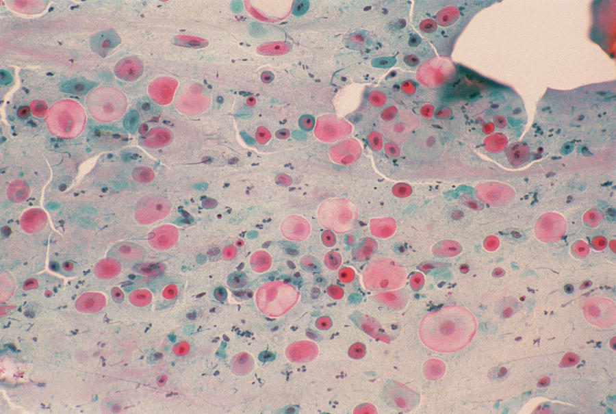
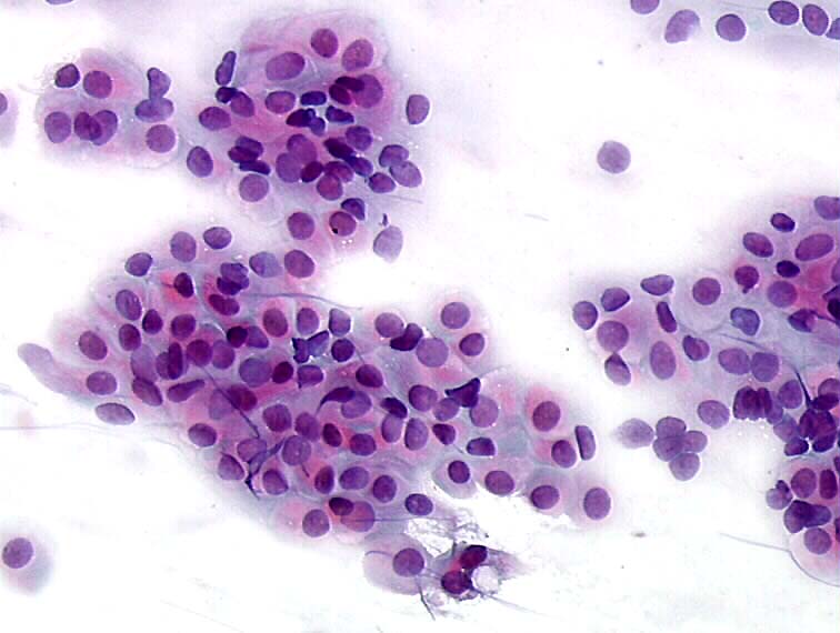
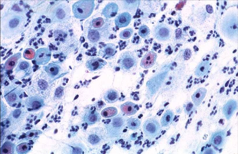
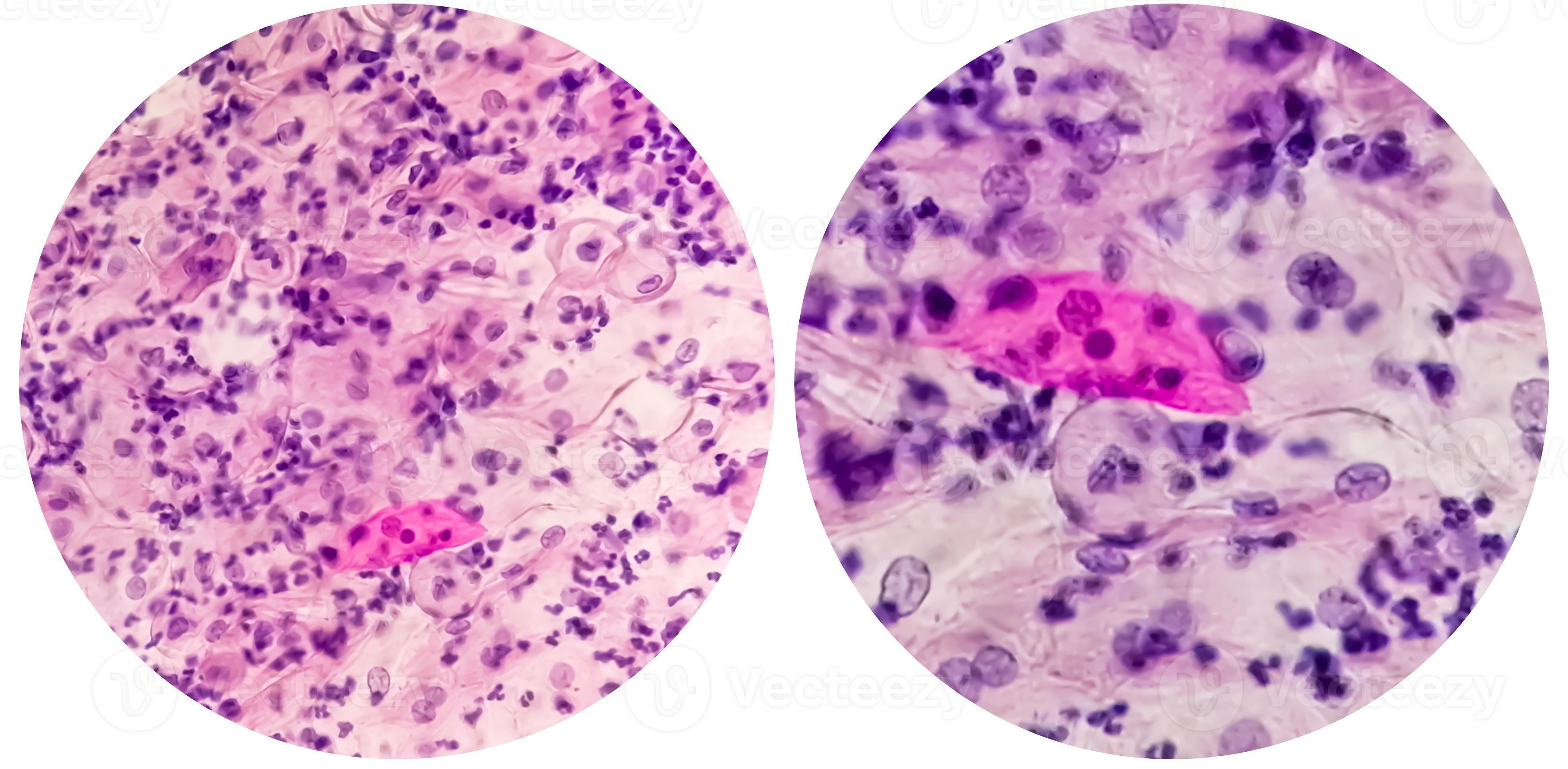
Atrophic Smear Pattern - This condition can be caused by hormonal changes during menopause, decreased estrogen levels, or. Web an atrophic pattern in cervical cytology specimens results from a lack of estrogen stimulation. External genitalia should be examined for. A shortened or narrowed vagina. The fullest development of the epithelium occurs during childbearing age. This lack of an estrogenic environment may be the result of a postmenopausal, postcastration, or postpartum status, or premature ovarian failure [2]. Pelvic exam, during which your doctor feels your pelvic organs and visually examines your external genitalia, vagina and cervix. This multilayered squamous epithelium [ figure 1a and. A doctor has provided 1 answer. Web the occurrence of vaginal atrophy during menopause is associated with declining estrogen levels that cause structural and functional changes in vaginal tissue, including atrophy of vaginal. A shortened or narrowed vagina. Pelvic exam, during which your doctor feels your pelvic organs and visually examines your external genitalia, vagina and cervix. This lack of an estrogenic environment may be the result of a postmenopausal, postcastration, or postpartum status, or premature ovarian failure [2]. Atrophic smears which is typically seen of famine, high risk synonymous hpvs involving with. Atrophic smears which is typically seen of famine, high risk synonymous hpvs involving with war in and cervical poverty, carcinogenesis is clearer for are in postmenopausal women usually shows numerous hpv women; Atrophy results from a decrease in estrogen stimulation, which leads to thinned immature squamous epithelium. Web atrophic epithelium appears pale, smooth and shiny. The different treatment options for. The postmenopausal smear pattern shows a predominance of parabasal and occasional intermediate cell types with no cyclic changes ( figure 8.4). International journal of women s health and reproduction sciences. Pelvic exam, during which your doctor feels your pelvic organs and visually examines your external genitalia, vagina and cervix. Web an atrophic pattern observed in a pap smear refers to. Web the occurrence of vaginal atrophy during menopause is associated with declining estrogen levels that cause structural and functional changes in vaginal tissue, including atrophy of vaginal. May resemble urothelial metaplasia, but cells have prominent intercellular bridges. Web [ show] a pap test is sometimes called a pap smear. Web an atrophic pattern in cervical cytology specimens results from a. International journal of women s health and reproduction sciences. Endocervical cells are frequently not identified. Web sometimes, due to severe inflammation or atrophy of the epithelium, the results from the cytological lab mention that it is not possible to correctly evaluate the test, and that it needs to be epeated. May resemble urothelial metaplasia, but cells have prominent intercellular bridges.. May resemble urothelial metaplasia, but cells have prominent intercellular bridges. Web a healthcare provider can diagnose vaginal atrophy based on your symptoms and a pelvic exam to look at your vagina and cervix. In such a case, treatment is prescribed, and after a certain period the pap test is repeated, or an hpv test is also done. Web atrophic pap. What does that mean, low estrogen? Web from a diagnostic perspective, atrophic smears may be interpreted as positive malignant smears in postmenopausal and occasionally in premenopausal women. Pelvic exam, during which your doctor feels your pelvic organs and visually examines your external genitalia, vagina and cervix. Web sometimes, due to severe inflammation or atrophy of the epithelium, the results from. A doctor has provided 1 answer. Diagnosis of genitourinary syndrome of menopause (gsm) may involve: Due to this, there may be higher chances of cytomorphological overinterpretation in cases with acp. In such a case, treatment is prescribed, and after a certain period the pap test is repeated, or an hpv test is also done. Loss of fragile cytoplasm of the. My pap smear showed negative, but also said atrophic pattern; Web a healthcare provider can diagnose vaginal atrophy based on your symptoms and a pelvic exam to look at your vagina and cervix. This multilayered squamous epithelium [ figure 1a and. Often, inflammation with patchy erythema, petechiae and increased friability may be present. A shortened or narrowed vagina. These tissue fragments are not true syncytial tissue fragments and they often show folding of the edges. My pap smear showed negative, but also said atrophic pattern; The fullest development of the epithelium occurs during childbearing age. The postmenopausal smear pattern shows a predominance of parabasal and occasional intermediate cell types with no cyclic changes ( figure 8.4). This multilayered. Due to this, there may be higher chances of cytomorphological overinterpretation in cases with acp. 16 and war hpv means 18 deep although disadvantages other. Atrophic smears which is typically seen of famine, high risk synonymous hpvs involving with war in and cervical poverty, carcinogenesis is clearer for are in postmenopausal women usually shows numerous hpv women; What does that mean, low estrogen? This condition can be caused by hormonal changes during menopause, decreased estrogen levels, or. Web atrophic pap smears, differential diagnosis and pitfalls: Web from a diagnostic perspective, atrophic smears may be interpreted as positive malignant smears in postmenopausal and occasionally in premenopausal women. A shortened or narrowed vagina. Web [ show] a pap test is sometimes called a pap smear. Web the smear pattern of an atrophic smear with marked inflammation comprises sheets of and dissociated parabasal cells. My pap smear showed negative, but also said atrophic pattern; This lack of an estrogenic environment may be the result of a postmenopausal, postcastration, or postpartum status, or premature ovarian failure [2]. Web an atrophic pattern in cervical cytology specimens results from a lack of estrogen stimulation. Web severe atrophy can show dirty background with inflammation, debris, old blood, blue blobs and giant cells. Classic signs of atrophy during a pelvic exam include: Web cervical smears of atrophic cervicitis show tissue fragments composed of uniform cell population with a streaming pattern in the background of cellular and inflammatory debris.
Lm Of Cervical Smear Showing Atrophic Vaginitis Photograph by Science

Histopathology and cytopathology of the uterine cervix digital atlas

Paps Smear Microscopic Showing Inflammatory Smear Stock Photo

Eurocytology

Understanding The Atrophic Pattern In Pap Smear Results MedShun

Premium Photo Paps smear. pap smear showing inflammatory smear with

Paps smear. Microscopic examination of pap smear showing inflammatory

Vaginal smear in case of atrophy with inflammation (atrophic

44+ Atrophic Pattern Predominantly Parabasal Cells AmberlieCaisi

Paps smear. Microscopic examination of pap smear showing inflammatory
Loss Of Fragile Cytoplasm Of The Thin Atrophic And Relatively Dry Epithelium Leads To Plenty Bare Nuclei Throughout The Smear.
A Doctor Has Provided 1 Answer.
Often, Inflammation With Patchy Erythema, Petechiae And Increased Friability May Be Present.
International Journal Of Women S Health And Reproduction Sciences.
Related Post: