Ana Pattern Centromere
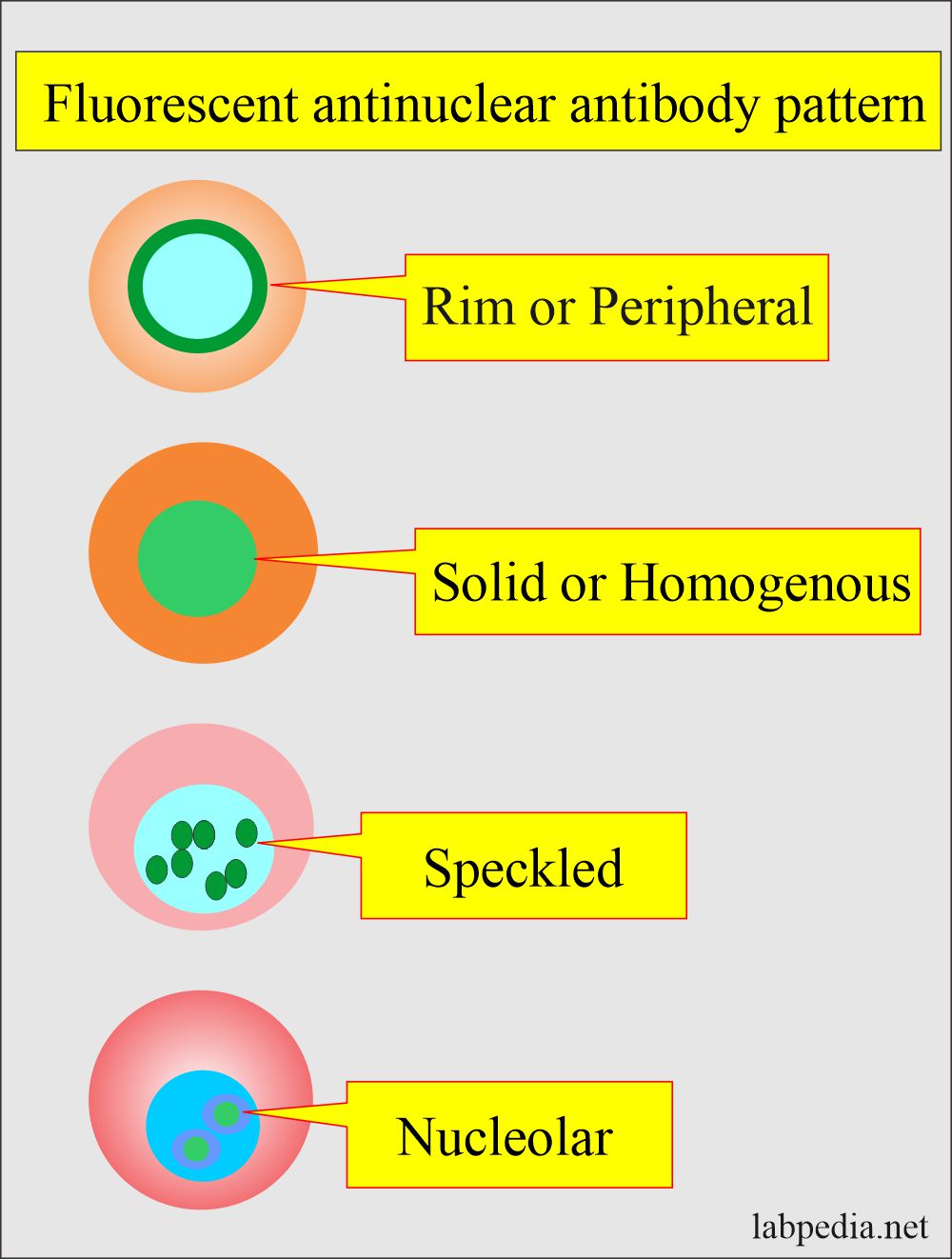
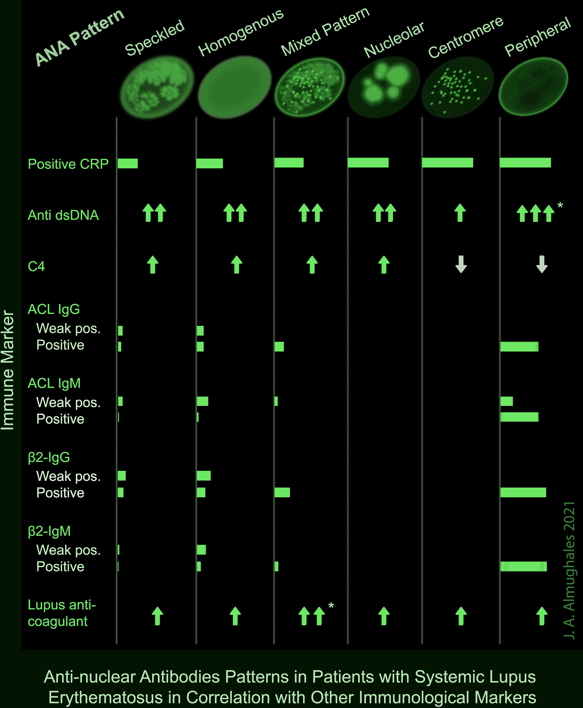
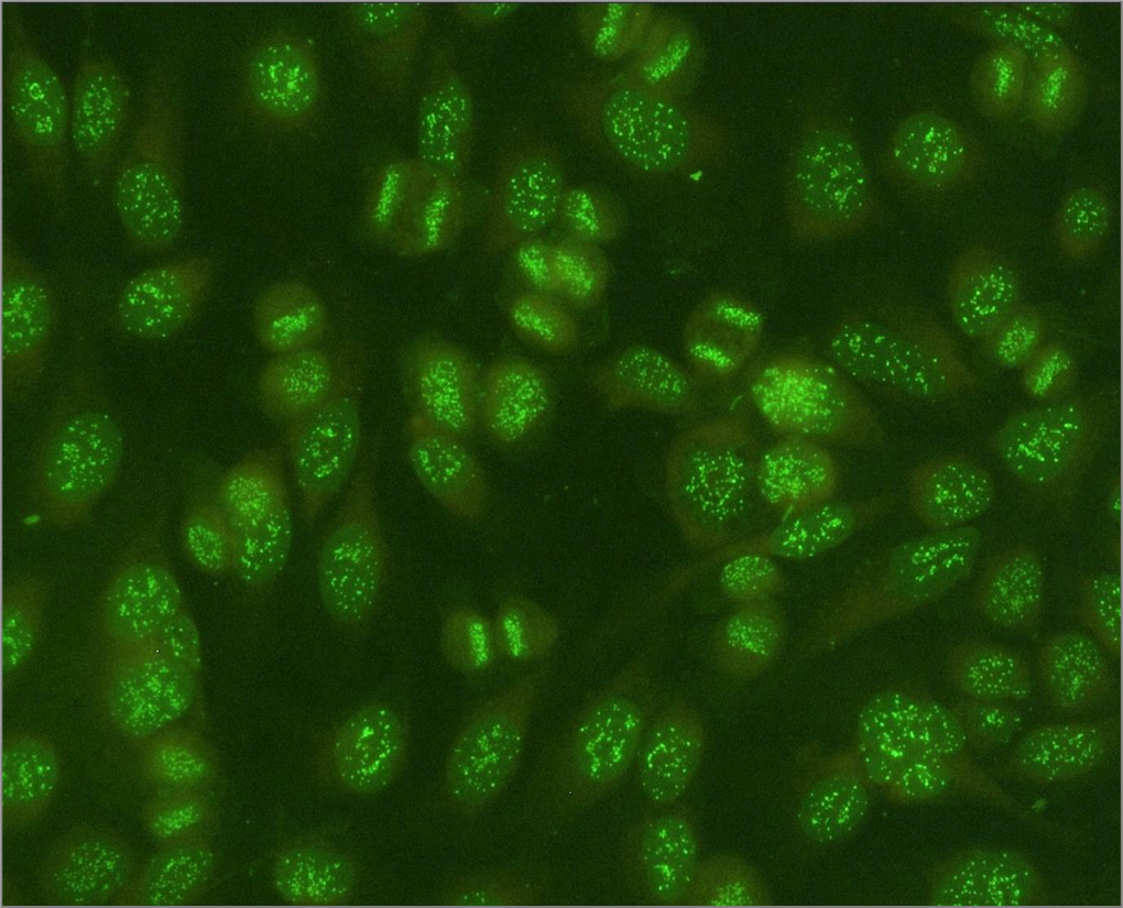
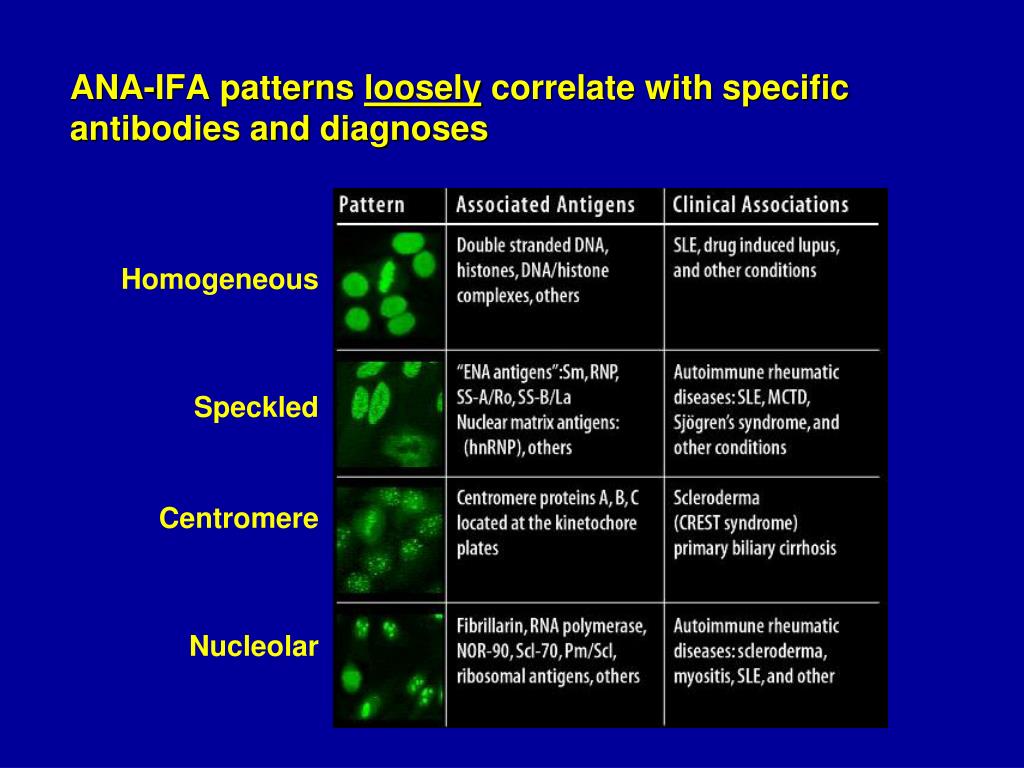
Ana Pattern Centromere - However, characteristics of aca in comparison with the other ana patterns and. Main antinuclear antibody patterns on immunofluorescence [1] Web my first ana panel came back positive for ana titer 1, 1:160 with a centromere pattern, nothing else came back positive.all symptoms still there and getting worse, now i've added night sweats and stomach cramps (that would wake me up in the night, found out that was coming from the plaquenil which has now been stopped) and. At times, laboratories testing ana also report a “pattern”. Web is the ana pattern suggestive of a specific disease? They may also be observed in other autoimmune diseases such as sjogren’s syndrome, rheumatoid arthritis, primary biliary cholangitis and overlap. Ana patterns are frequently interpreted incorrectly and thus i would question the result. Patterns that are reported include, homogeneous, speckled, centromere, and others. A membranous pattern may show antibodies to membrane proteins. Web ana shows up on iif assays as a fluorescent pattern in cells that are fixed to a slide. The prevalence and clinical significance of uncommon or rare patterns, particularly those directed at the mitotic spindle apparatus (msa), are not. Scleroderma 70 antibody aka topoisomerase 1. T is true that a centromere ana pattern is associated with crest. Patterns that are reported include, homogeneous, speckled, centromere, and others. Web iif patterns correlate to specific ana subtypes, and pattern recognition. Nuclear speckled pattern with striking variability in intensity with the strongest staining in g2 phase and weakest/negative staining in g1. Main antinuclear antibody patterns on immunofluorescence [1] The pattern refers to the distribution of staining produced by autoantibodies reacting with antigens in the cells. Seen in people with limited scleroderma. A membranous pattern may show antibodies to membrane proteins. Web ana antibody patterns were described to be as peripheral, speckled, homogenous, nucleolar, and centromere patterns. A centromere staining pattern means the ana staining is present along the chromosomes. Nuclear speckled pattern with striking variability in intensity with the strongest staining in g2 phase and weakest/negative staining in g1. Confirms centromere pattern on ana and seen in those with limited. The level or titer and the pattern. Web an antinuclear antibody test is a blood test that looks for certain kinds of antibodies in your body. A nucleolar staining pattern means ana staining is present around the. At times, laboratories testing ana also report a “pattern”. Web ana shows up on iif assays as a fluorescent pattern in cells that. Web a speckled pattern may indicate various diseases, including lupus and sjögren’s syndrome. Some, but not all labs will report a titre above 1:160 as positive. The nucleus is essentially the “command centre” or “brain” of any cell in the body. It’s also called an ana or fana (fluorescent antinuclear antibody) test. Homogenous fluorescence pattern typically suggests antibodies directed at. A nucleolar staining pattern means ana staining is present around the. Nuclear speckled pattern with striking variability in intensity with the strongest staining in g2 phase and weakest/negative staining in g1. Web ana test results are most often reported in 2 parts: Web is the ana pattern suggestive of a specific disease? Homogenous fluorescence pattern typically suggests antibodies directed at. The nucleus is essentially the “command centre” or “brain” of any cell in the body. Therefore, the pattern can be further investigated under a microscope. T is true that a centromere ana pattern is associated with crest. Although there are some overlaps, different patterns can be associated with certain autoimmune diseases. Web nuclear speckled pattern with striking variability in intensity. A centromere pattern may indicate scleroderma. Scleroderma 70 antibody aka topoisomerase 1. Seen in people with limited scleroderma. Homogenous fluorescence pattern typically suggests antibodies directed at dsdna, histones, or nucleosomes. Some, but not all labs will report a titre above 1:160 as positive. The centromeres are positive only in prometaphase and metaphase, revealing multiple aligned small and faint dots. For this test, we use a. Although there are some overlaps, different patterns can be associated with certain autoimmune diseases. A centromere staining pattern means the ana staining is present along the chromosomes. A membranous pattern may show antibodies to membrane proteins. Titres are reported in ratios, most often 1:40, 1:80, 1:160, 1:320, and 1:640. Medical records of patients with suspected aild who had positive cytoplasmic ana patterns between february 2017 and november 2019 were retrospectively reviewed for clinical, laboratory, and immunological data. Web my first ana panel came back positive for ana titer 1, 1:160 with a centromere pattern, nothing else. The centromeres are positive only in prometaphase and metaphase, revealing multiple aligned small and faint dots. A membranous pattern may show antibodies to membrane proteins. Ideally, one should provide the most complete definition of the pattern (expert. Individuals with high titers of these antibodies may lack serious organ involvement, being classified as having undifferentiated connective tissue disease (uctd) or. Scleroderma 70 antibody aka topoisomerase 1. T is true that a centromere ana pattern is associated with crest. Patterns that are reported include, homogeneous, speckled, centromere, and others. Homogenous fluorescence pattern typically suggests antibodies directed at dsdna, histones, or nucleosomes. Web nuclear speckled pattern with striking variability in intensity with the strongest staining in g2 phase and weakest/negative staining in g1. Web ana shows up on iif assays as a fluorescent pattern in cells that are fixed to a slide. A nucleolar staining pattern means ana staining is present around the. Nuclear speckled pattern with striking variability in intensity with the strongest staining in g2 phase and weakest/negative staining in g1. Main antinuclear antibody patterns on immunofluorescence [1] The prevalence and clinical significance of uncommon or rare patterns, particularly those directed at the mitotic spindle apparatus (msa), are not. Medical records of patients with suspected aild who had positive cytoplasmic ana patterns between february 2017 and november 2019 were retrospectively reviewed for clinical, laboratory, and immunological data. It’s also called an ana or fana (fluorescent antinuclear antibody) test.
Centromere staining pattern in indirect fluorescence Download

Centromere ANA, AC3 from homepage of International consensus of ANA

Ana Test Patterns
Antinuclear Factor (ANF), Antinuclear Antibody (ANA) and Its
Frontiers AntiNuclear Antibodies Patterns in Patients With Systemic
Antinuclear antibodies (ANA) positive control centromere pattern

Common ANA patterns by IIF a, negative sample; b, homogeneous; c
PPT Choosing the Correct ANA Technology for your Laboratory
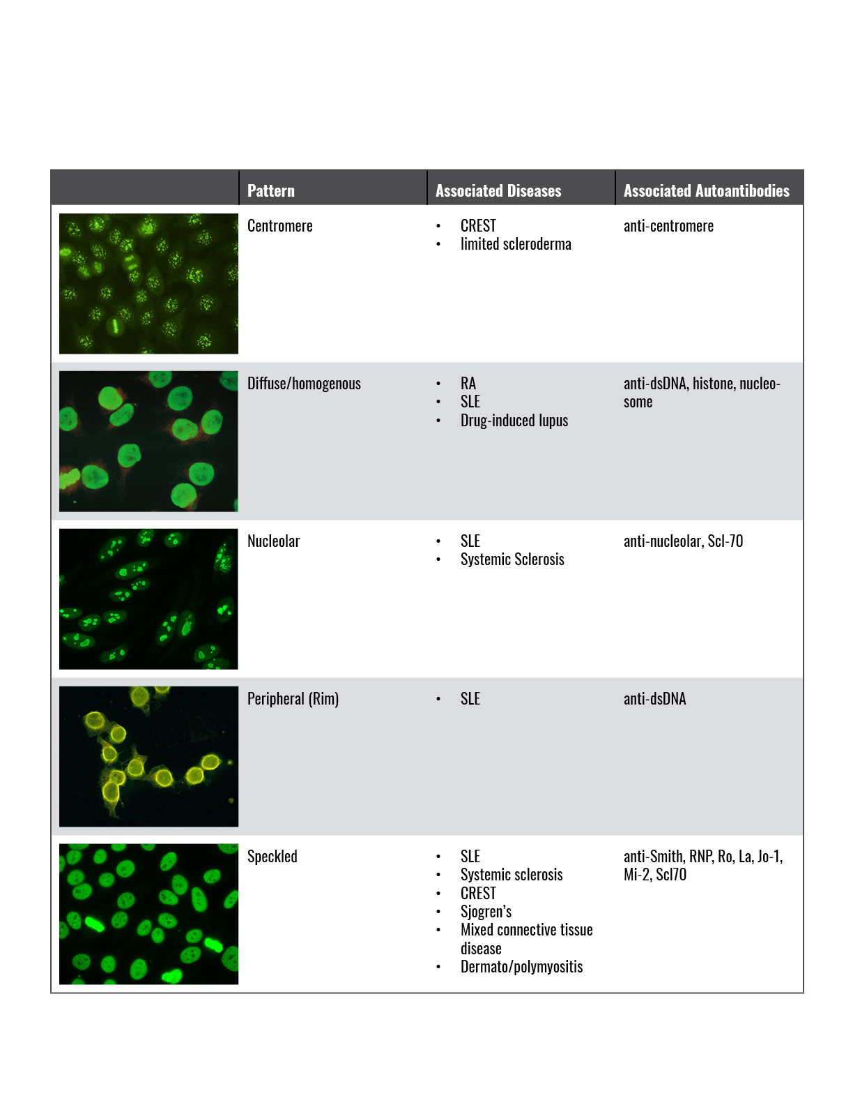
Ana Test Patterns

Anticentromere antibodies target
Some, But Not All Labs Will Report A Titre Above 1:160 As Positive.
However, Characteristics Of Aca In Comparison With The Other Ana Patterns And.
Web An Antinuclear Antibody Test Is A Blood Test That Looks For Certain Kinds Of Antibodies In Your Body.
Web Ana Test Results Are Most Often Reported In 2 Parts:
Related Post: