Ana Homogenous Pattern
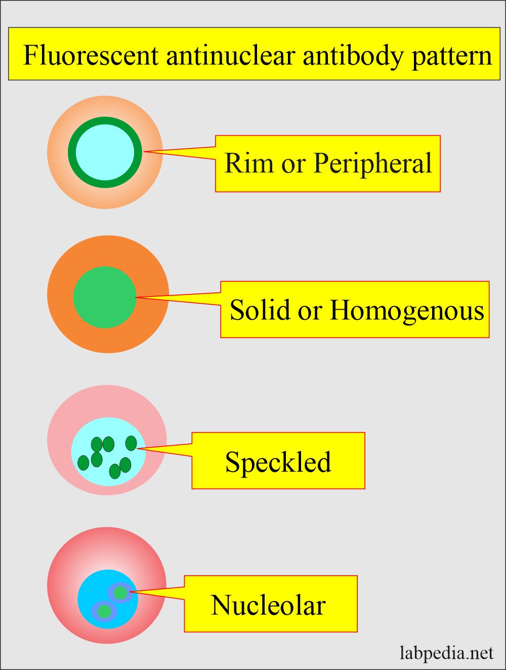
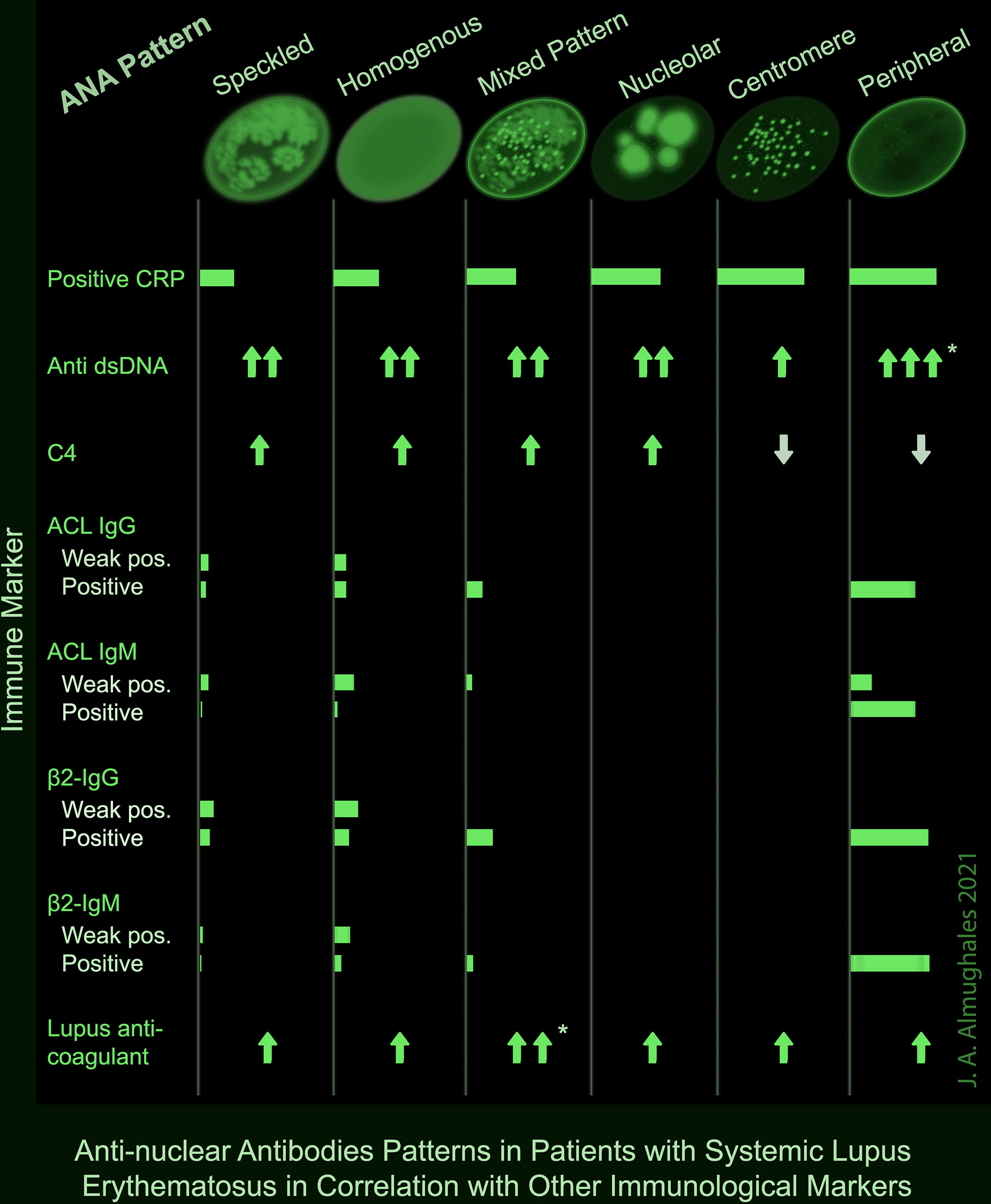
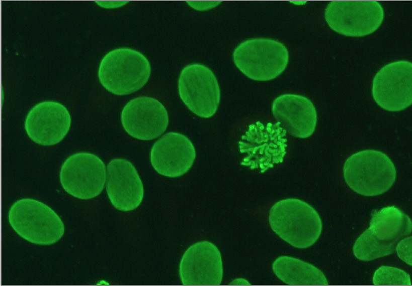
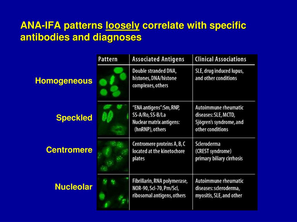
Ana Homogenous Pattern - The commonly recognized patterns include: The quantity of ana in the serum (intensity) and, when the ana is positive, the pattern of antibody binding to the nucleus (staining pattern). Web an ana test detects antinuclear antibodies (ana) in your blood. A peripheral pattern indicates that fluorescence occurs at the edges of the nucleus in a shaggy appearance; Fine and coarse speckles of ana staining are seen throughout the nucleus. The entire nucleus is stained with ana. Your immune system normally makes antibodies to help you fight infection. A homogenous (diffuse) pattern appears as total nuclear fluorescence and is common in people with systemic lupus. Interphase cells show homogeneous nuclear staining while mitotic cells show staining of the condensed chromosome regions. Anas are typically classified into two groups, antibodies to dna and histones and antibodies to nuclear material. Fine and coarse speckles of ana staining are seen throughout the nucleus. Ana is usually measured as 0 to 4+ or as a titer (the number of times a blood sample can be diluted and still be positive). The most common antibody test is called the antinuclear antibody (ana) test. Doctors may order an ana test if you have signs. A speckled staining pattern means fine, coarse speckles of ana are present. If your doctor thinks you might have lupus, they may ask you to take blood tests to check for antibodies in your blood. The quantity of ana in the serum (intensity) and, when the ana is positive, the pattern of antibody binding to the nucleus (staining pattern). Web. When active, usually a homogenous pattern on ana or less commonly speckled, rim, or nucleolar when present in. The level or titer and the pattern. Although nearly all patients with sle have positive ana titers, most patients with a positive titer do not have. The entire nucleus is stained with ana. The quantity of ana in the serum (intensity) and,. Web the classical nuclear patterns are speckled, homogeneous, nucleolar and centromere. Web the antinuclear antibody (ana) test. Interphase cells show homogeneous nuclear staining while mitotic cells show staining of the condensed chromosome regions. Web the pattern of the ana test can give information about the type of autoimmune disease present and the appropriate treatment program. Although nearly all patients with. Web the presence of ana with a homogeneous & speckled (hs) pattern was significantly associated with the absence of cancer ( < 0.01). Your immune system normally makes antibodies to help you fight infection. A homogenous staining pattern means the entire nucleus is stained with ana. Web the pattern of the ana test can give information about the type of. Antibodies that attack healthy proteins within the cell nucleus are called antinuclear antibodies (anas). Doctors may order an ana test if you have signs or symptoms. Web an ana test detects antinuclear antibodies (ana) in your blood. Ana is usually measured as 0 to 4+ or as a titer (the number of times a blood sample can be diluted and. Antibodies that attack healthy proteins within the cell nucleus are called antinuclear antibodies (anas). Web the antinuclear antibody (ana) test. It’s also called an ana or fana (fluorescent antinuclear antibody) test. In contrast, antinuclear antibodies often attack your body's own. The most common antibody test is called the antinuclear antibody (ana) test. Web an ana test detects antinuclear antibodies (ana) in your blood. A titer (a measure of how much ana is in the blood) and a pattern (where the ana was detected in the cells). The most common antibody test is called the antinuclear antibody (ana) test. Web the classical nuclear patterns are speckled, homogeneous, nucleolar and centromere. This pattern is. Web the presence of ana with a homogeneous & speckled (hs) pattern was significantly associated with the absence of cancer ( < 0.01). A homogenous pattern can mean any autoimmune disease but more specifically, lupus or sjögren’s syndrome. This is the most common pattern and can be seen with any autoimmune disease. The level or titer and the pattern. If. Web homogenous and/or nuclear rim (peripheral) pattern correlates with antibody to native dna and deoxynucleoprotein and bears correlation with sle, sle activity, and lupus nephritis. Web a positive nuclear staining result will usually come back with a more detailed staining pattern, such as speckled (fig. Anas are typically classified into two groups, antibodies to dna and histones and antibodies to. Fine and coarse speckles of ana staining are seen throughout the nucleus. Anas are typically classified into two groups, antibodies to dna and histones and antibodies to nuclear material. Web the classical nuclear patterns are speckled, homogeneous, nucleolar and centromere. The entire nucleus is stained with ana. Web homogenous and/or nuclear rim (peripheral) pattern correlates with antibody to native dna and deoxynucleoprotein and bears correlation with sle, sle activity, and lupus nephritis. This is the most common pattern and can be seen with any autoimmune disease. A speckled staining pattern means fine, coarse speckles of ana are present. The commonly recognized patterns include: If your doctor thinks you might have lupus, they may ask you to take blood tests to check for antibodies in your blood. Web ana titers and patterns can vary between laboratory testing sites due to variations in the methodology used. Interphase cells show homogeneous nuclear staining while mitotic cells show staining of the condensed chromosome regions. Your immune system normally makes antibodies to help you fight infection. It’s also called an ana or fana (fluorescent antinuclear antibody) test. The level or titer and the pattern. Doctors may order an ana test if you have signs or symptoms. A speckled pattern is also found in lupus.
ANA Patterns

Ana Pattern Homogeneous Chumado

Common ANA patterns by IIF a, negative sample; b, homogeneous; c

6. IFA pattern Homogeneous ANA pattern YouTube

Antinuclear Factor (ANF), Antinuclear Antibody (ANA) and Its

DFS70 antibodies biomarkers for the exclusion of ANAassociated

Frontiers AntiNuclear Antibodies Patterns in Patients With Systemic

ANA Patterns

Antinuclear antibodies (ANA) homogeneous pattern positive control

Homogeneous Ana Pattern Pagswa
Homogenous (Diffuse) Pattern Suggests Sle Or Other Connective Tissue Diseases.
Web Antinuclear Antibodies (Ana) Refer To An Autoantibody Directed At Material Within The Nucleus Of A Cell.
The Most Common Antibody Test Is Called The Antinuclear Antibody (Ana) Test.
A Titer (A Measure Of How Much Ana Is In The Blood) And A Pattern (Where The Ana Was Detected In The Cells).
Related Post: