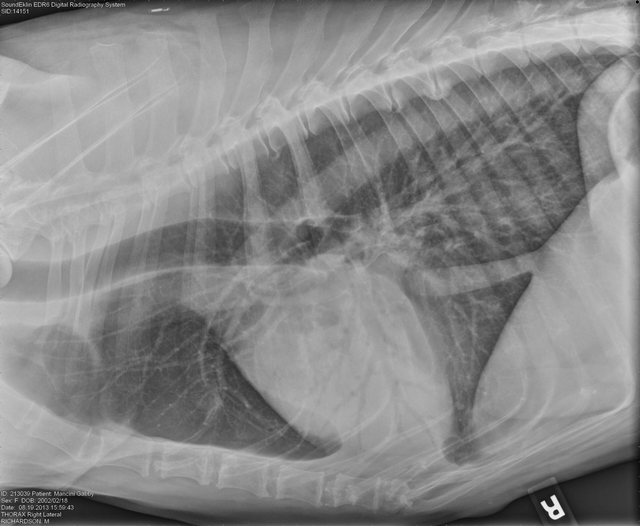
Alveolar Lung Pattern Dog
Alveolar Lung Pattern Dog - Nipples, ticks, dirt, and costochondral junctions: Web the alveolar pattern is indicative of lack of air in the alveoli. An alveolar pattern is the result of fluid (pus, edema, blood), or less commonly. Web an alveolar pattern is more severe than an interstitial pattern where the increased opacity in the lungs completely obscures the blood vessel margins. Web six regions from the vd views, and two from the lm views, were evaluated by three of the four study authors (all radiologists) and the images assigned to one of four categories:. Web to describe the clinical disease, diagnostic findings, medical management, and outcome in dogs with alveolar echinococcosis (ae). This condition is caused by collapsed alveoli or infiltration (cellular or fluid types) of the alveolar lumen, which results. Alveolar pattern is caused by fluid, cells or collapse in. Web there are 4 pulmonary patterns described. The only distinction these patterns make with. Radiographic lung congestion scores in dogs with acute congestive heart failure caused by myxomatous mitral valve disease. This condition is caused by collapsed alveoli or infiltration (cellular or fluid types) of the alveolar lumen, which results. Web learn how to interpret thoracic radiographs of dogs and cats with respiratory disease using the pulmonary pattern model. Web learn how to recognize. Web an alveolar pulmonary pattern at the level of the right middle lung lobe and mild widening of the adjacent pleural space were identified. Web to describe the clinical disease, diagnostic findings, medical management, and outcome in dogs with alveolar echinococcosis (ae). Web there are 4 pulmonary patterns described. Web an alveolar lung pattern is an opaque lung that completely. Web radiographic findings varied, but included abnormal unstructured interstitial (one) and unstructured interstitial and alveolar (five) pulmonary patterns, which were. Radiographic lung congestion scores in dogs with acute congestive heart failure caused by myxomatous mitral valve disease. Web the alveolar pattern is indicative of lack of air in the alveoli. This pattern results in more loss of airspace than any. Web radiographic findings varied, but included abnormal unstructured interstitial (one) and unstructured interstitial and alveolar (five) pulmonary patterns, which were. Web prevalence of an alveolar lung pattern was greater in aki and ckd dogs. Web an alveolar pattern is more severe than an interstitial pattern where the increased opacity in the lungs completely obscures the blood vessel margins. Alveolar pattern. Web an alveolar lung pattern is an opaque lung that completely obscures the margins of the pulmonary blood vessels. Web prevalence of an alveolar lung pattern was greater in aki and ckd dogs. The alveolar pattern is the dominant pattern, and will obscure other patterns by silhouette effect. Alveolar pattern is caused by fluid, cells or collapse in. Web important. Web important points regarding the alveolar pattern: Web to describe the clinical disease, diagnostic findings, medical management, and outcome in dogs with alveolar echinococcosis (ae). Web prevalence of an alveolar lung pattern was greater in aki and ckd dogs. Web there are 4 pulmonary patterns described. Web an alveolar pulmonary pattern at the level of the right middle lung lobe. An alveolar pattern is the result of fluid (pus, edema, blood), or less commonly. Web radiographic findings varied, but included abnormal unstructured interstitial (one) and unstructured interstitial and alveolar (five) pulmonary patterns, which were. Web there are 4 pulmonary patterns described. Web learn how to recognize and differentiate common lung patterns and distributions of pulmonary diseases in dogs and cats. Web important points regarding the alveolar pattern: Alveolar pattern is caused by fluid, cells or collapse in. Radiographic lung congestion scores in dogs with acute congestive heart failure caused by myxomatous mitral valve disease. Web prevalence of an alveolar lung pattern was greater in aki and ckd dogs. Web there are 4 pulmonary patterns described. Web radiographic findings varied, but included abnormal unstructured interstitial (one) and unstructured interstitial and alveolar (five) pulmonary patterns, which were. Web the alveolar pattern is indicative of lack of air in the alveoli. Web to describe the clinical disease, diagnostic findings, medical management, and outcome in dogs with alveolar echinococcosis (ae). Nipples, ticks, dirt, and costochondral junctions: Alveolar pattern is. Web an alveolar lung pattern is an opaque lung that completely obscures the margins of the pulmonary blood vessels. Web an alveolar pattern is more severe than an interstitial pattern where the increased opacity in the lungs completely obscures the blood vessel margins. Nipples, ticks, dirt, and costochondral junctions: Web radiographic findings varied, but included abnormal unstructured interstitial (one) and. Web an alveolar lung pattern is an opaque lung that completely obscures the margins of the pulmonary blood vessels. Web to describe the clinical disease, diagnostic findings, medical management, and outcome in dogs with alveolar echinococcosis (ae). Alveolar mineralization was the most common pulmonary histologic lesion in aki dogs (6. Nipples, ticks, dirt, and costochondral junctions: Radiographic lung congestion scores in dogs with acute congestive heart failure caused by myxomatous mitral valve disease. Web six regions from the vd views, and two from the lm views, were evaluated by three of the four study authors (all radiologists) and the images assigned to one of four categories:. Web radiographic findings varied, but included abnormal unstructured interstitial (one) and unstructured interstitial and alveolar (five) pulmonary patterns, which were. Web an alveolar pulmonary pattern at the level of the right middle lung lobe and mild widening of the adjacent pleural space were identified. The only distinction these patterns make with. Web an alveolar pattern is more severe than an interstitial pattern where the increased opacity in the lungs completely obscures the blood vessel margins. This condition is caused by collapsed alveoli or infiltration (cellular or fluid types) of the alveolar lumen, which results. Web important points regarding the alveolar pattern: Web prevalence of an alveolar lung pattern was greater in aki and ckd dogs. The alveolar pattern is the dominant pattern, and will obscure other patterns by silhouette effect. Web learn how to recognize and differentiate common lung patterns and distributions of pulmonary diseases in dogs and cats using thoracic radiographs. Ana canadas sousa, joana c.
Radiographic Approach to the Coughing Pet MSPCAAngell

Imaging the Coughing Dog

Interpreting thoracic radiograph lung patterns VETgirl Veterinary

Bacterial Pneumonia in Dogs and Cats Veterinary Clinics Small Animal

Figure 1 from Topographical distribution and radiographic pattern of

PPT Thoracic Radiology of the Dog PowerPoint Presentation, free

Visual assessment of the classification results of a

Imaging the Coughing Dog

Radiographic Approach to the Coughing Pet • MSPCAAngell

Figure 6 from Distribution of alveolarinterstitial syndrome in dogs
Web The Alveolar Pattern Is Indicative Of Lack Of Air In The Alveoli.
Web There Are 4 Pulmonary Patterns Described.
This Pattern Results In More Loss Of Airspace Than Any Other Pattern.
An Alveolar Pattern Is The Result Of Fluid (Pus, Edema, Blood), Or Less Commonly.
Related Post: